Application
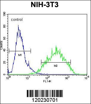
Flow cytometric analysis of NIH-3T3 cells (right histogram) compared to a negative control cell (left histogram).FITC-conjugated goat-anti-rabbit secondary antibodies were used for the analysis.Application
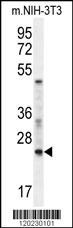
Western blot analysis in mouse NIH-3T3 cell line lysates (35ug/lane).Application
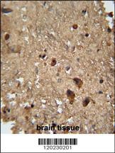
RAN antibody immunohistochemistry analysis in formalin fixed and paraffin embedded human brain tissue followed by peroxidase conjugation of the secondary antibody and DAB staining.| Product Name | RAN Antibody |
|---|---|
| Antibody Type | Primary Antibodies |
| Product description | RAN (ras-related nuclear protein) is a small GTP binding protein belonging to the RAS superfamily that is essential for the translocation of RNA and proteins through the nuclear pore complex. The RAN protein is also involved in control of DNA synthesis and cell cycle progression. Nuclear localization of RAN requires the presence of regulator of chromosome condensation 1 (RCC1). Mutations in RAN disrupt DNA synthesis. Because of its many functions, it is likely that RAN interacts with several other proteins. RAN regulates formation and organization of the microtubule network independently of its role in the nucleus-cytosol exchange of macromolecules. RAN could be a key signaling molecule regulating microtubule polymerization during mitosis. RCC1 generates a high local concentration of RAN-GTP around chromatin which, in turn, induces the local nucleation of microtubules. RAN is an androgen receptor (AR) coactivator that binds differentially with different lengths of polyglutamine within the androgen receptor. Polyglutamine repeat expansion in the AR is linked to Kennedy's disease (X-linked spinal and bulbar muscular atrophy). RAN coactivation of the AR diminishes with polyglutamine expansion within the AR, and this weak coactivation may lead to partial androgen insensitivity during the development of Kennedy's disease.1) Kim, J.S., et al. Mol. Carcinog. 49(10):913-921(2010) |
| Immunogen | This RAN antibody is generated from rabbits immunized with a KLH conjugated synthetic peptide between 112-140 amino acids from the Central region of human RAN. |
| Clonality | Polyclonal |
|---|---|
| Isotype | Ig |
| Host Species | Rabbit |
| Tested Applications | FACSIHC-PWB |
| For WB starting dilution is: 1:1000 For IHC-P starting dilution is: 1:50~100 For FACS starting dilution is: 1:10~50 | |
| Species Reactivity | HumanMouse |
| Concentration | 1mg/ml |
| Purification | Affinity purified |
| Gene Symbol | RAN |
|---|---|
| Alternative Names | GTP-binding nuclear protein Ran Androgen receptor-associated protein 24 GTPase Ran Ras-like protein TC4 Ras-related nuclear protein RAN ARA24 |
| Molecular Weight(MW) | 24 kDa |
| Sequence Similarities | Predicted species reactivity based on immunogen sequence: Bovine |
Application
Flow cytometric analysis of NIH-3T3 cells (right histogram) compared to a negative control cell (left histogram).FITC-conjugated goat-anti-rabbit secondary antibodies were used for the analysis.
Application
Western blot analysis in mouse NIH-3T3 cell line lysates (35ug/lane).
Application
RAN antibody immunohistochemistry analysis in formalin fixed and paraffin embedded human brain tissue followed by peroxidase conjugation of the secondary antibody and DAB staining.| Application Notes | For WB starting dilution is: 1:1000 For IHC-P starting dilution is: 1:50~100 For FACS starting dilution is: 1:10~50 |
|---|
| Form | Liquid |
|---|---|
| Storage Instructions | Store at 4˚C for three months and -20˚C, stable for up to one year. As with all antibodies care should be taken to avoid repeated freeze thaw cycles. Antibodies should not be exposed to prolonged high temperatures. |
| Storage Buffer | Supplied in PBS with 0.09% (W/V) sodium azide. |


