Application
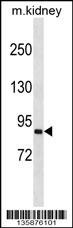
Western blot analysis in mouse kidney tissue lysates (35ug/lane).Application
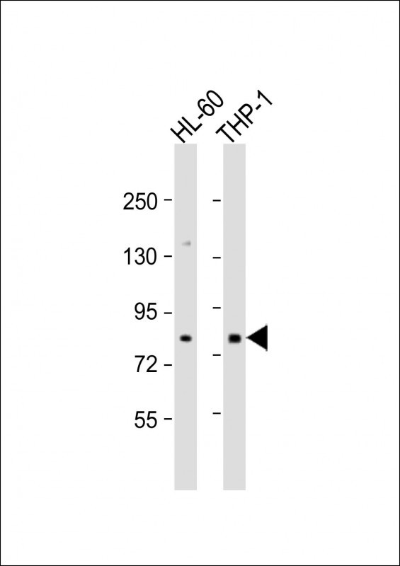
Western Blot at 1:500 dilution Lane 1: HL-60 whole cell lysate Lane 2: THP-1 whole cell lysate Lysates/proteins at 20 ug per lane.| Product Name | SEMA4A Antibody |
|---|---|
| Antibody Type | Primary Antibodies |
| Product description | This gene encodes a member of the semaphorin family of soluble and transmembrane proteins. Semaphorins are involved in numerous functions, including axon guidance, morphogenesis, carcinogenesis, and immunomodulation. The encoded protein is a single-pass type I membrane protein containing an immunoglobulin-like C2-type domain, a PSI domain and a sema domain. It inhibits axonal extension by providing local signals to specify territories inaccessible for growing axons. It is an activator of T-cell-mediated immunity and suppresses vascular endothelial growth factor (VEGF)-mediated endothelial cell migration and proliferation in vitro and angiogenesis in vivo. Mutations in this gene are associated with retinal degenerative diseases including retinitis pigmentosa type 35 (RP35) and cone-rod dystrophy type 10 (CORD10). Multiple alternatively spliced transcript variants encoding different isoforms have been identified.1) Davila, S., et al. Genes Immun. 11(3):232-238(2010) |
| Immunogen | This SEMA4A antibody is generated from rabbits immunized with a KLH conjugated synthetic peptide between 151-180 amino acids from the N-terminal region of human SEMA4A. |
| Clonality | Polyclonal |
|---|---|
| Isotype | Ig |
| Host Species | Rabbit |
| Tested Applications | WB |
| For WB starting dilution is: 1:500 | |
| Species Reactivity | HumanMouse |
| Concentration | 1mg/ml |
| Purification | Affinity purified |
| Gene Symbol | SEMA4A |
|---|---|
| Alternative Names | Semaphorin-4A Semaphorin-B Sema B SEMA4A SEMAB SEMB |
| Molecular Weight(MW) | 84 kDa |
Application
Western blot analysis in mouse kidney tissue lysates (35ug/lane).
Application
Western Blot at 1:500 dilution Lane 1: HL-60 whole cell lysate Lane 2: THP-1 whole cell lysate Lysates/proteins at 20 ug per lane.| Application Notes | For WB starting dilution is: 1:500 |
|---|
| Form | Liquid |
|---|---|
| Storage Instructions | Store at 4˚C for three months and -20˚C, stable for up to one year. As with all antibodies care should be taken to avoid repeated freeze thaw cycles. Antibodies should not be exposed to prolonged high temperatures. |
| Storage Buffer | Supplied in PBS with 0.09% (W/V) sodium azide. |


