Application
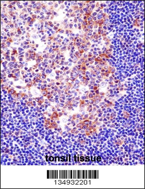
SPI1 Antibody immunohistochemistry analysis in formalin fixed and paraffin embedded human tonsil tissue followed by peroxidase conjugation of the secondary antibody and DAB staining.Application
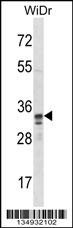
Western blot analysis in WiDr cell line lysates (35ug/lane).Application
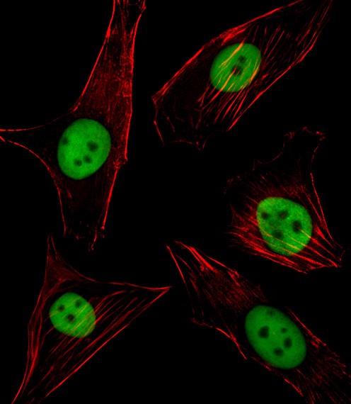
Fluorescent image of NIH/3T3 cell stained with SPI1 Antibody .NIH/3T3 cells were fixed with 4% PFA (20 min), permeabilized with Triton X-100 (0.1%, 10 min), then incubated with SPI1 primary antibody (1:25). For secondary antibody, Alexa Fluor 488 conjugated donkey anti-rabbit antibody (green) was used (1:400).Cytoplasmic actin was counterstained with Alexa Fluor 555 (red) conjugated Phalloidin (7u| Product Name | SPI1 Antibody |
|---|---|
| Antibody Type | Primary Antibodies |
| Antigen Alias | Transcription factor PU1, 31 kDa-transforming protein, SPI1 |
| Product description | This gene encodes an ETS-domain transcription factor that activates gene expression during myeloid and B-lymphoid cell development. The nuclear protein binds to a purine-rich sequence known as the PU-box found near the promoters of target genes, and regulates their expression in coordination with other transcription factors and cofactors. The protein can also regulate alternative splicing of target genes. Multiple transcript variants encoding different isoforms have been found for this gene. [provided by RefSeq].1) Rimmele, P., et al. Cancer Res. 70(17):6757-6766(2010) |
| Immunogen | This SPI1 antibody is generated from rabbits immunized with a KLH conjugated synthetic peptide between 217-246 amino acids from the C-terminal region of human SPI1. |
| Clonality | Polyclonal |
|---|---|
| Isotype | Ig |
| Host Species | Rabbit |
| Tested Applications | IFIHC-PWB |
| For IF starting dilution is: 1:10~50 For WB starting dilution is: 1:1000 For IHC-P starting dilution is: 1:10~50: |
|
| Species Reactivity | Human |
| Concentration | 1mg/ml |
| Purification | Affinity purified |
| Gene Symbol | SPI1 |
|---|---|
| Alternative Names | Transcription factor PU1 31 kDa-transforming protein SPI1 |
| Molecular Weight(MW) | 31 kDa |
| Sequence Similarities | Predicted species reactivity based on immunogen sequence: Mouse |
Application
SPI1 Antibody immunohistochemistry analysis in formalin fixed and paraffin embedded human tonsil tissue followed by peroxidase conjugation of the secondary antibody and DAB staining.
Application
Western blot analysis in WiDr cell line lysates (35ug/lane).
Application
Fluorescent image of NIH/3T3 cell stained with SPI1 Antibody .NIH/3T3 cells were fixed with 4% PFA (20 min), permeabilized with Triton X-100 (0.1%, 10 min), then incubated with SPI1 primary antibody (1:25). For secondary antibody, Alexa Fluor 488 conjugated donkey anti-rabbit antibody (green) was used (1:400).Cytoplasmic actin was counterstained with Alexa Fluor 555 (red) conjugated Phalloidin (7u| Application Notes | For IF starting dilution is: 1:10~50 For WB starting dilution is: 1:1000 For IHC-P starting dilution is: 1:10~50: |
|---|
| Form | Liquid |
|---|---|
| Storage Instructions | Store at 4˚C for three months and -20˚C, stable for up to one year. As with all antibodies care should be taken to avoid repeated freeze thaw cycles. Antibodies should not be exposed to prolonged high temperatures. |
| Storage Buffer | Supplied in PBS with 0.09% (W/V) sodium azide. |


