Application
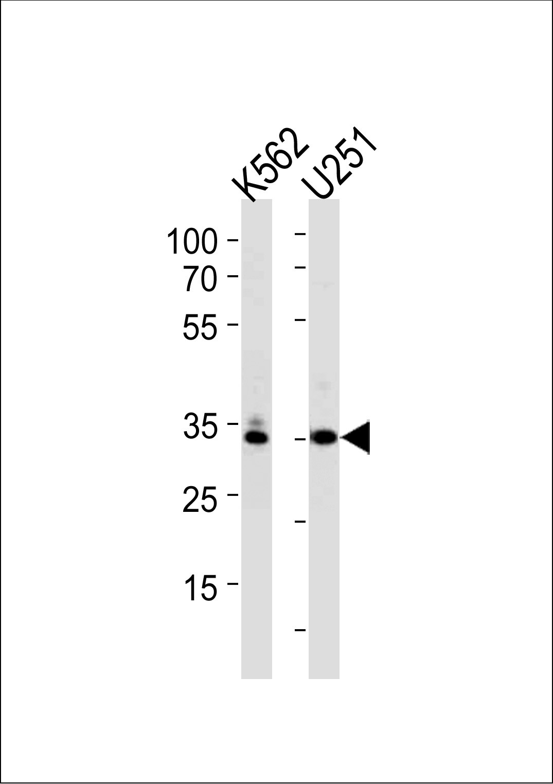
Western blot analysis in K562,U251 cell line lysates (35ug/lane).Application
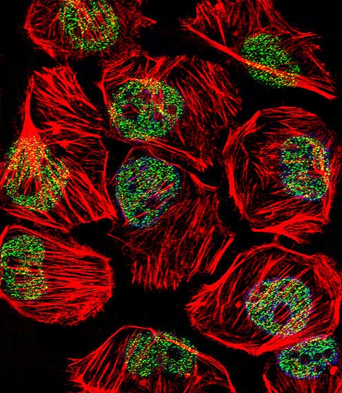
Fluorescent confocal image of U251 cell stained with TBP Antibody.U251 cells were fixed with 4% PFA (20 min), permeabilized with Triton X-100 (0.1%, 10 min), then incubated with TBP primary antibody (1:25). For secondary antibody, Alexa Fluor 488 conjugated donkey anti-rabbit antibody (green) was used (1:400).Cytoplasmic actin was counterstained with Alexa Fluor 555 (red) conjugated Phalloidin (7uApplication
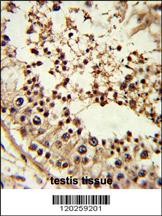
Formalin-fixed and paraffin-embedded human testis tissue reacted with TBP Antibody , which was peroxidase-conjugated to the secondary antibody, followed by DAB staining.Application
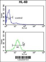
Flow cytometry analysis of HL-60 cells (bottom histogram) compared to a negative control cell (top histogram).FITC-conjugated goat-anti-rabbit secondary antibodies were used for the analysis.| Product Name | TBP Antibody |
|---|---|
| Antibody Type | Primary Antibodies |
| Antigen Alias | TATA-box-binding protein, TATA sequence-binding protein, TATA-binding factor, TATA-box factor, Transcription initiation factor TFIID TBP subunit, TBP, GTF2D1, TF2D, TFIID |
| Product description | Initiation of transcription by RNA polymerase II requires the activities of more than 70 polypeptides. The protein that coordinates these activities is transcription factor IID (TFIID), which binds to the core promoter to position the polymerase properly, serves as the scaffold for assembly of the remainder of the transcription complex, and acts as a channel for regulatory signals. TFIID is composed of the TATA-binding protein (TBP) and a group of evolutionarily conserved proteins known as TBP-associated factors or TAFs. TAFs may participate in basal transcription, serve as coactivators, function in promoter recognition or modify general transcription factors (GTFs) to facilitate complex assembly and transcription initiation. A distinctive feature of TBP is a long string of glutamines in the N-terminal. This region of the protein modulates the DNA binding activity of the C terminus, and modulation of DNA binding affects the rate of transcription complex formation and initiation of transcription. Mutations that expand the number of CAG repeats encoding this polyglutamine tract, and thus increase the length of the polyglutamine string, are associated with spinocerebellar ataxia 17, a neurodegenerative disorder classified as a polyglutamine disease.1) Ohi,K., Genes Brain Behav.(4), 473-480 (2009) |
| Immunogen | This TBP antibody is generated from rabbits immunized with a KLH conjugated synthetic peptide between 210-239 amino acids from the Central region of human TBP. |
| Clonality | Polyclonal |
|---|---|
| Isotype | Ig |
| Host Species | Rabbit |
| Tested Applications | FACSIFIHC-PWB |
| For WB starting dilution is: 1:1000 For IF starting dilution is: 1:10~50 For IHC-P starting dilution is: 1:50~100 For FACS starting dilution is: 1:10~50: |
|
| Species Reactivity | Human |
| Concentration | 1mg/ml |
| Gene Symbol | TBP |
|---|---|
| Alternative Names | TATA-box-binding protein TATA sequence-binding protein TATA-binding factor TATA-box factor Transcription initiation factor TFIID TBP subunit TBP GTF2D1 TF2D TFIID |
| Molecular Weight(MW) | 38 kDa |
| Sequence Similarities | Predicted species reactivity based on immunogen sequence: Zebrafish |
Application
Western blot analysis in K562,U251 cell line lysates (35ug/lane).
Application
Fluorescent confocal image of U251 cell stained with TBP Antibody.U251 cells were fixed with 4% PFA (20 min), permeabilized with Triton X-100 (0.1%, 10 min), then incubated with TBP primary antibody (1:25). For secondary antibody, Alexa Fluor 488 conjugated donkey anti-rabbit antibody (green) was used (1:400).Cytoplasmic actin was counterstained with Alexa Fluor 555 (red) conjugated Phalloidin (7u
Application
Formalin-fixed and paraffin-embedded human testis tissue reacted with TBP Antibody , which was peroxidase-conjugated to the secondary antibody, followed by DAB staining.
Application
Flow cytometry analysis of HL-60 cells (bottom histogram) compared to a negative control cell (top histogram).FITC-conjugated goat-anti-rabbit secondary antibodies were used for the analysis.| Application Notes | For WB starting dilution is: 1:1000 For IF starting dilution is: 1:10~50 For IHC-P starting dilution is: 1:50~100 For FACS starting dilution is: 1:10~50: |
|---|
| Form | Liquid |
|---|---|
| Storage Instructions | Store at 4˚C for three months and -20˚C, stable for up to one year. As with all antibodies care should be taken to avoid repeated freeze thaw cycles. Antibodies should not be exposed to prolonged high temperatures. |
| Storage Buffer | Supplied in PBS with 0.09% (W/V) sodium azide. |


