Application
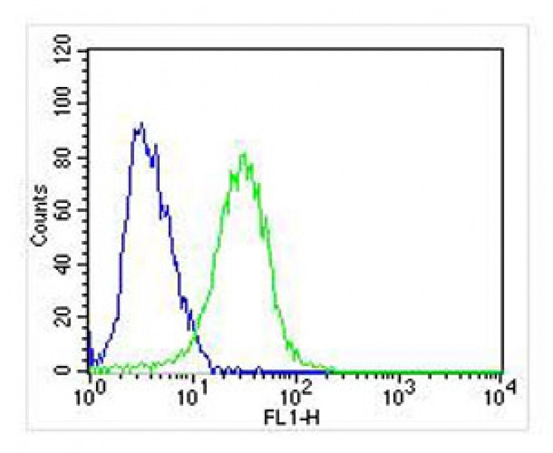
Overlay histogram showing SH-SY5Y cells stained with Antibody (green line). The cells were fixed with 2% paraformaldehyde (10 min). The cells were then icubated in 2% bovine serum albumin to block non-specific protein-protein interactions followed by the antibody (1:25 dilution) for 60 min at 37ºC. The secondary antibody used was Goat-Anti-Rabbit IgG, DyLight 488 Conjugated Highly Cross-Adsorbed(OApplication
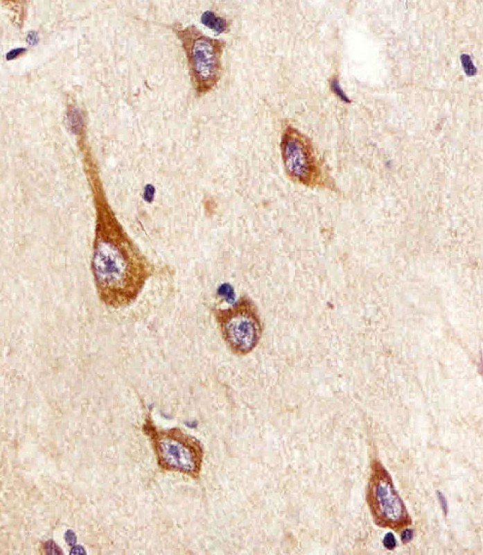
Antibody staining TrkA in human brain tissue sections by Immunohistochemistry (IHC-P - paraformaldehyde-fixed, paraffin-embedded sections).Application
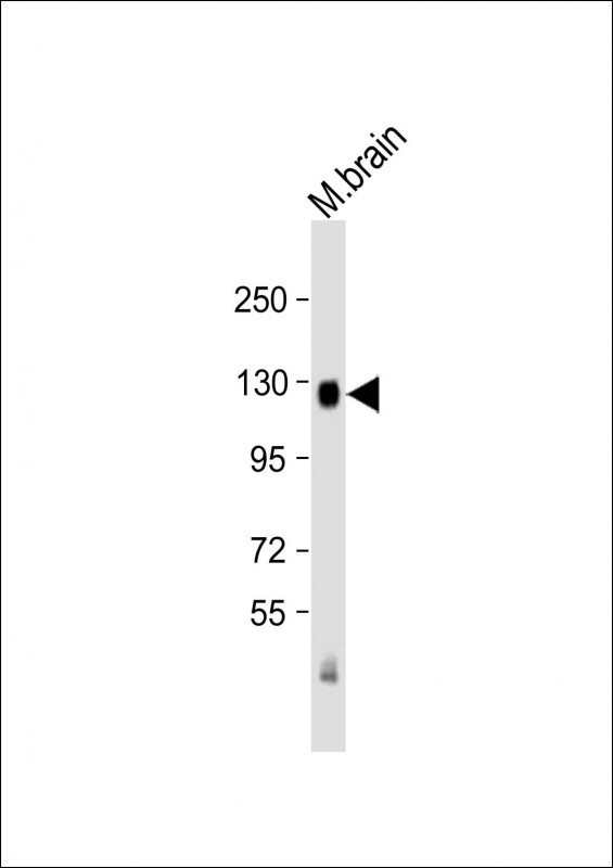
Western Blot at 1:2000 dilution + mouse brain lysate Lysates/proteins at 20 ug per lane.Application
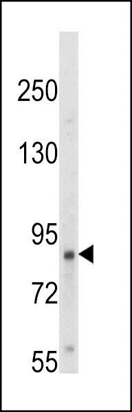
Western blot analysis of hTrkA-pY791 in HepG2 cell line lysates (35ug/lane)Application
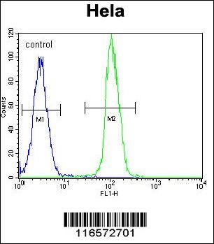
Flow cytometric analysis of Hela cells (right histogram) compared to a negative control cell (left histogram).FITC-conjugated goat-anti-rabbit secondary antibodies were used for the analysis.Application
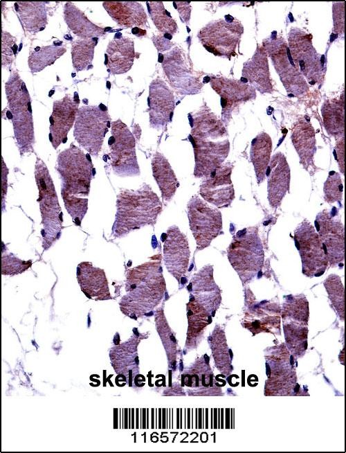
TrkA-pY791 Antibody immunohistochemistry analysis in formalin fixed and paraffin embedded human skeletal muscle followed by peroxidase conjugation of the secondary antibody and DAB staining.| Product Name | TrkA-pY791 Antibody |
|---|---|
| Antibody Type | Primary Antibodies |
| Antigen Alias | High affinity nerve growth factor receptor, Neurotrophic tyrosine kinase receptor type 1, TRK1-transforming tyrosine kinase protein, Tropomyosin-related kinase A, Tyrosine kinase receptor, Tyrosine kinase receptor A, Trk-A, gp140trk, p140-TrkA, NTRK1, MTC, |
| Product description | TRKA (also known as NTRK1)is a member of the neurotrophic tyrosine kinase receptor (NTKR) family. This kinase is a membrane-bound receptor that, upon neurotrophin binding, phosphorylates itself and members of the MAPK pathway. The presence of this kinase leads to cell differentiation and may play a role in specifying sensory neuron subtypes. Mutations in the TRKA gene have been associated with congenital insensitivity to pain, anhidrosis, self-mutilating behavior, mental retardation and cancer.1) Tokusashi, Y., et al., Int. J. Cancer 114(1):39-45 (2005). |
| Immunogen | This TrkA antibody is generated from rabbits immunized with a KLH conjugated synthetic peptide between 769-796 amino acids from human TrkA. |
| Clonality | Polyclonal |
|---|---|
| Isotype | Ig |
| Host Species | Rabbit |
| Tested Applications | FACSIHC-PWB |
| For FACS starting dilution is: 1:25 For IHC-P starting dilution is: 1:25 For WB starting dilution is: 1:1000: |
|
| Species Reactivity | HumanMouse |
| Concentration | 1mg/ml |
| Purification | Affinity purified |
| Gene Symbol | NTRK1 |
|---|---|
| Alternative Names | High affinity nerve growth factor receptor Neurotrophic tyrosine kinase receptor type 1 TRK1-transforming tyrosine kinase protein Tropomyosin-related kinase A Tyrosine kinase receptor Tyrosine kinase receptor A Trk-A gp140trk p140-TrkA NTRK1 MTC |
| Molecular Weight(MW) | 87 kDa |
Application
Overlay histogram showing SH-SY5Y cells stained with Antibody (green line). The cells were fixed with 2% paraformaldehyde (10 min). The cells were then icubated in 2% bovine serum albumin to block non-specific protein-protein interactions followed by the antibody (1:25 dilution) for 60 min at 37ºC. The secondary antibody used was Goat-Anti-Rabbit IgG, DyLight 488 Conjugated Highly Cross-Adsorbed(O
Application
Antibody staining TrkA in human brain tissue sections by Immunohistochemistry (IHC-P - paraformaldehyde-fixed, paraffin-embedded sections).
Application
Western Blot at 1:2000 dilution + mouse brain lysate Lysates/proteins at 20 ug per lane.
Application
Western blot analysis of hTrkA-pY791 in HepG2 cell line lysates (35ug/lane)
Application
Flow cytometric analysis of Hela cells (right histogram) compared to a negative control cell (left histogram).FITC-conjugated goat-anti-rabbit secondary antibodies were used for the analysis.
Application
TrkA-pY791 Antibody immunohistochemistry analysis in formalin fixed and paraffin embedded human skeletal muscle followed by peroxidase conjugation of the secondary antibody and DAB staining.| Application Notes | For FACS starting dilution is: 1:25 For IHC-P starting dilution is: 1:25 For WB starting dilution is: 1:1000: |
|---|
| Form | Liquid |
|---|---|
| Storage Instructions | Store at 4˚C for three months and -20˚C, stable for up to one year. As with all antibodies care should be taken to avoid repeated freeze thaw cycles. Antibodies should not be exposed to prolonged high temperatures. |
| Storage Buffer | Supplied in PBS with 0.09% (W/V) sodium azide. |


 Data sheet for OM294146