Application
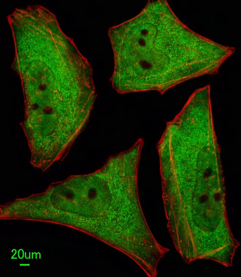
Immunofluorescent analysis of Hela cells, using Ubiquilin1 Antibody . Antibody was diluted at 1:100 dilution. Alexa Fluor 488-conjugated goat anti-rabbit lgG at 1:400 dilution was used as the secondary antibody (green). Cytoplasmic actin was counterstained with Dylight Fluor 554 (red) conjugated Phalloidin (red).Application
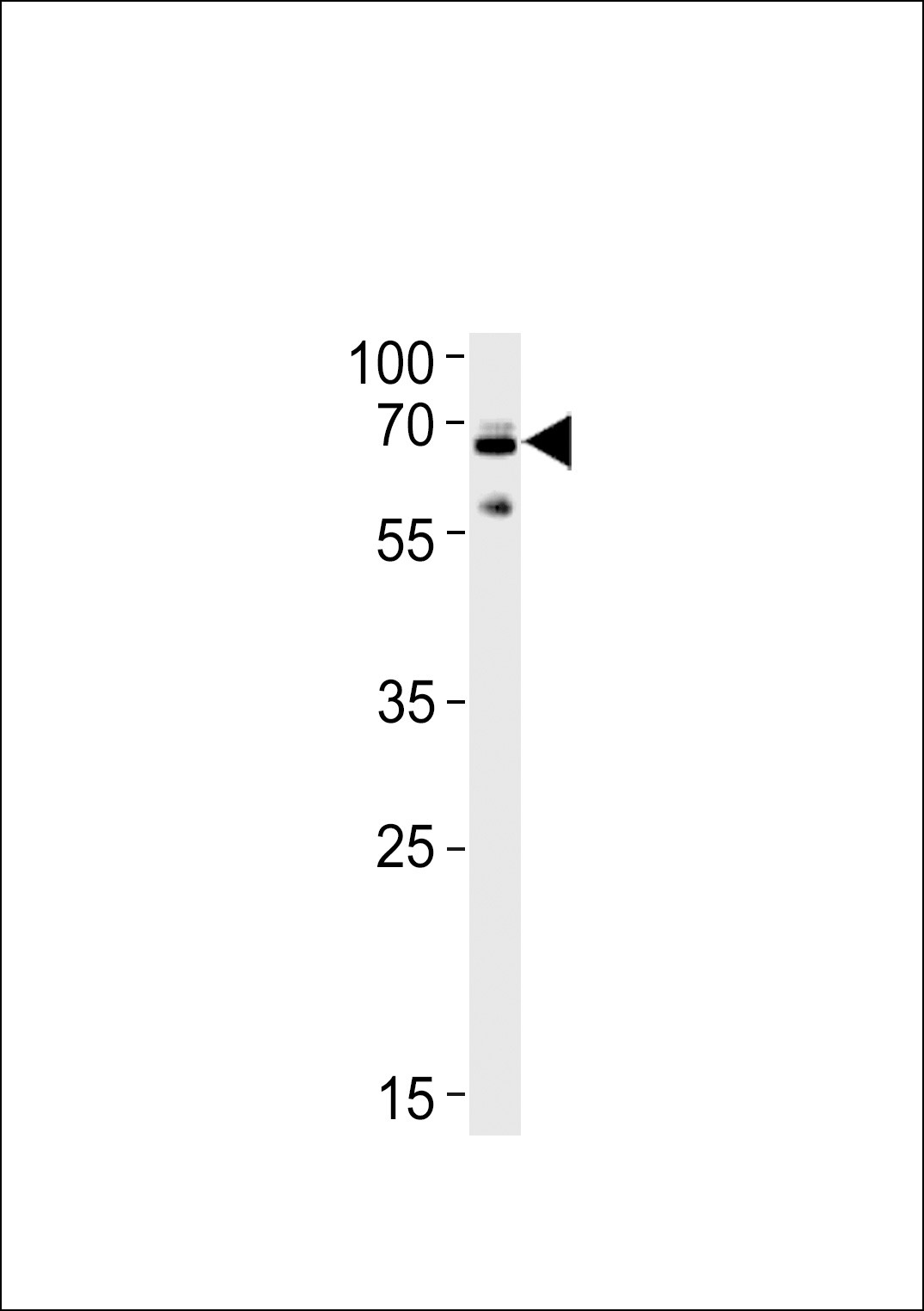
Western blot analysis in SH-SY5Y cell line lysates (35ug/lane).This demonstrates the Ubiquilin1 antibody detected the Ubiquilin1 protein (arrow).Application
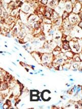
Formalin-fixed and paraffin-embedded human cancer tissue reacted with the primary antibody, which was peroxidase-conjugated to the secondary antibody, followed by AEC staining. BC = breast carcinoma; HC = hepatocarcinoma.| Product Name | Ubiquilin1 Antibody |
|---|---|
| Antibody Type | Primary Antibodies |
| Antigen Alias | Ubiquilin-1, Protein linking IAP with cytoskeleton 1, PLIC-1, hPLIC-1, UBQLN1, DA41, PLIC1 |
| Product description | Ubiquilin 1 (UBQLN1), also known as DA41, was isolated from an adult rat lung cDNA library, and encodes a cellular protein that associates with DAN.1 DAN expression is reduced in rat fibroblast 3Y1 cells transformed with mouse sarcoma virus and in rodent fibroblasts transformed with a variety of oncogenes. The DAN-DA41 interaction is mediated through the N-terminal domain and a cysteine-knot region of DAN. Human DA41 encodes a 589-amino acid protein with 86% amino acid sequence identity with rat protein.2 DA41 expression is regulated in a cell cycle-dependent manner. PLIC1 and PLIC2 (UBQLN2) are homologs of the mouse Plics (proteins linking integrin-associated protein (IAP) and cytoskeleton) and the yeast Dsk2 protein. PLIC1, also called UBQLN1, shares 72% amino acid identity with PLIC2,3 Two motifs are conserved in the mammalian PLICs and yeast Dsk2, an N-terminal ubiquitin-like (UBL) domain and a C-terminal ubiquitin-associated (UBA) domain. Unlike ubiquitin, the UBL domain of the PLICs does not have a diglycine motif in its C terminus. The UBA domain is present in multiple enzyme classes of the ubiquitination machinery. Human PLICs associate with both proteasomes and ubiquitin ligases in large complexes. Overexpression of PLICs impairs the in vivo degradation of 2 unrelated ubiquitin-dependent proteasome substrates, p53 and I-kappa-B-alpha (NFKBIA), but not a ubiquitin-independent substrate. PLICs may link the ubiquitination machinery to the proteasome to affect in vivo protein degradation. The DA41 gene maps to chromosome 9q21.2-q21.3, a position overlapping a candidate tumor suppressor locus for bladder cancer.21) Massey, L.K., et al., J. Alzheimers Dis. 6(1):79-92 (2004). Gao, L., et al., J. Virol. 77(7):4149-4159 (2003). Mah, A.L., et al., J. Cell Biol. 151(4):847-862 (2000). Kleijnen, M.F., et al., Mol. Cell 6(2):409-419 (2000). Hanaoka, E., et al., J. Hum. Genet. 45(3):188-191 (2000). |
| Immunogen | This Ubiquilin1 antibody is generated from rabbits immunized with a KLH conjugated synthetic peptide between 296-326 amino acids from the Central region of human Ubiquilin1. |
| Clonality | Polyclonal |
|---|---|
| Isotype | Ig |
| Host Species | Rabbit |
| Tested Applications | IFIHC-PWB |
| For IF starting dilution is: 1:100 For WB starting dilution is: 1:1000 For IHC-P starting dilution is: 1:50~100: |
|
| Species Reactivity | HumanMouse |
| Concentration | 1mg/ml |
| Gene Symbol | UBQLN1 |
|---|---|
| Alternative Names | Ubiquilin-1 Protein linking IAP with cytoskeleton 1 PLIC-1 hPLIC-1 UBQLN1 DA41 PLIC1 |
| Molecular Weight(MW) | 63 kDa |
| Sequence Similarities | Predicted species reactivity based on immunogen sequence: Rat |
Application
Immunofluorescent analysis of Hela cells, using Ubiquilin1 Antibody . Antibody was diluted at 1:100 dilution. Alexa Fluor 488-conjugated goat anti-rabbit lgG at 1:400 dilution was used as the secondary antibody (green). Cytoplasmic actin was counterstained with Dylight Fluor 554 (red) conjugated Phalloidin (red).
Application
Western blot analysis in SH-SY5Y cell line lysates (35ug/lane).This demonstrates the Ubiquilin1 antibody detected the Ubiquilin1 protein (arrow).
Application
Formalin-fixed and paraffin-embedded human cancer tissue reacted with the primary antibody, which was peroxidase-conjugated to the secondary antibody, followed by AEC staining. BC = breast carcinoma; HC = hepatocarcinoma.| Application Notes | For IF starting dilution is: 1:100 For WB starting dilution is: 1:1000 For IHC-P starting dilution is: 1:50~100: |
|---|
| Form | Liquid |
|---|---|
| Storage Instructions | Store at 4˚C for three months and -20˚C, stable for up to one year. As with all antibodies care should be taken to avoid repeated freeze thaw cycles. Antibodies should not be exposed to prolonged high temperatures. |
| Storage Buffer | Supplied in PBS with 0.09% (W/V) sodium azide. |


