
Application
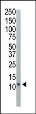
Western blot analysis of anti-Ubiquitin Pab in HeLa cell lysate. Ubiquitin was detected using purified Pab.Application
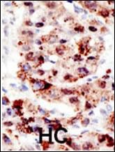
Formalin-fixed and paraffin-embedded human cancer tissue reacted with the primary antibody, which was peroxidase-conjugated to the secondary antibody, followed by AEC staining. BC = breast carcinoma; HC = hepatocarcinoma.Application
Characterization of FOXP2 Isoforms. FOXP2.10t was detected with an antibody to the N-terminal XpressTM tag or FOXP2 (green) and counterstained with antibodies to the aggresome marker ubiquitin (red). Nuclei are marked by DAPI staining (blue). Ubiquitin co-localizes with FOXP2.10t aggregates suggesting that these cellular bodies represent aggresomes.WB
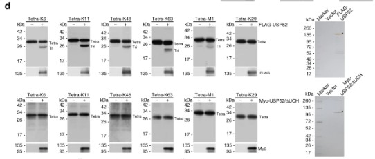
d In vitro deubiquitination assays with different types of ubiquitin linkages (1 μg) and HeLa cells-purified full-length USP52 (1 μg) or UCH domain deficient USP52 (USP52/∆UCH, 1 μg) with high salt and detergent buffer. After 4 h of incubation, the cleavage effect was examined by western blotting with antibody against ubiquitin. The asterisk indicates the recombinant protein stained by silver staining.| Product Name | Ubiquitin Antibody |
|---|---|
| Antibody Type | Primary Antibodies |
| Product description | This gene encodes ubiquitin, one of the most conserved proteins known. Ubiquitin is required for ATP-dependent, nonlysosomal intracellular protein degradation of abnormal proteins and normal proteins with a rapid turnover. Ubiquitin is covalently bound to proteins to be degraded, and presumably labels these proteins for degradation. Ubiquitin also binds to histone H2A in actively transcribed regions but does not cause histone H2A degradation, suggesting that ubiquitin is also involved in regulation of gene expression. This gene consists of three direct repeats of the ubiquitin coding sequence with no spacer sequence. Consequently, the protein is expressed as a polyubiquitin precursor with a final amino acid after the last repeat. Aberrant form of this protein has been noticed in patients with Alzheimer's and Down syndrome.1) Chan, Y.L., et al., Biochem. Biophys. Res. Commun. 215(2):682-690 (1995). |
| Immunogen | This Ubiquitin antibody is generated from rabbits immunized with a KLH conjugated synthetic peptide between 36-66 amino acids of human Ubiquitin. |
| Modification | Acetylated |
| Clonality | Polyclonal |
|---|---|
| Isotype | Ig |
| Host Species | Rabbit |
| Tested Applications | IFIHC-PWB |
| For WB starting dilution is: 1:1000 For IHC-P starting dilution is: 1:50~200 For IF starting dilution is: 1:100~500: |
|
| Species Reactivity | Human |
| Concentration | 1mg/ml |
| Gene Symbol | UBC |
|---|---|
| Alternative Names | Polyubiquitin-C Ubiquitin UBC |
| Molecular Weight(MW) | 77 kDa |
| Sequence Similarities | Predicted species reactivity based on immunogen sequence: Mouse |
Application
Western blot analysis of anti-Ubiquitin Pab in HeLa cell lysate. Ubiquitin was detected using purified Pab.
Application
Formalin-fixed and paraffin-embedded human cancer tissue reacted with the primary antibody, which was peroxidase-conjugated to the secondary antibody, followed by AEC staining. BC = breast carcinoma; HC = hepatocarcinoma.
Application
Characterization of FOXP2 Isoforms. FOXP2.10t was detected with an antibody to the N-terminal XpressTM tag or FOXP2 (green) and counterstained with antibodies to the aggresome marker ubiquitin (red). Nuclei are marked by DAPI staining (blue). Ubiquitin co-localizes with FOXP2.10t aggregates suggesting that these cellular bodies represent aggresomes.
WB
d In vitro deubiquitination assays with different types of ubiquitin linkages (1 μg) and HeLa cells-purified full-length USP52 (1 μg) or UCH domain deficient USP52 (USP52/∆UCH, 1 μg) with high salt and detergent buffer. After 4 h of incubation, the cleavage effect was examined by western blotting with antibody against ubiquitin. The asterisk indicates the recombinant protein stained by silver staining.| Application Notes | For WB starting dilution is: 1:1000 For IHC-P starting dilution is: 1:50~200 For IF starting dilution is: 1:100~500: |
|---|
| Form | Liquid |
|---|---|
| Storage Instructions | Store at 4˚C for three months and -20˚C, stable for up to one year. As with all antibodies care should be taken to avoid repeated freeze thaw cycles. Antibodies should not be exposed to prolonged high temperatures. |
| Storage Buffer | Supplied in PBS with 0.09% (W/V) sodium azide. |


