


Application
Fig1: Western blot analysis of KLF4 on different lysates with Rabbit anti-KLF4 antibody at 1/2,000 dilution. Lane 1: 293 cell lysate, Lane 2: NCCIT cell lysate, Lysates/proteins at 10 µg/Lane. Exposure time: 1 minute 30 seconds; 10% SDS-PAGE gel. Proteins were transferred to a PVDF membrane and blocked with 5% NFDM/TBST for 1 hour at room temperature. The primary antibody at 1/2,000 dilution was used in 5% NFDM/TBST at room temperature for 2 hours. Goat Anti-Rabbit IgG - HRP Secondary Antibody at 1:100,000 dilution was used for 1 hour at room temperature.Application
Fig2: ICC staining KLF4 in Hela cells (red). The nuclear counter stain is DAPI (blue). Cells were fixed in paraformaldehyde, permeabilised with 0.25% Triton X100/PBS.Application
Fig3: ICC staining KLF4 in PC-3M cells (red). The nuclear counter stain is DAPI (blue). Cells were fixed in paraformaldehyde, permeabilised with 0.25% Triton X100/PBS.Application
Fig4: ICC staining KLF4 in 293T cells (red). The nuclear counter stain is DAPI (blue). Cells were fixed in paraformaldehyde, permeabilised with 0.25% Triton X100/PBS.Application
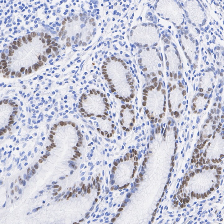
Fig5: Species: Human, Tissue: Stomach, Sample: Paraffin-embedded section.Application
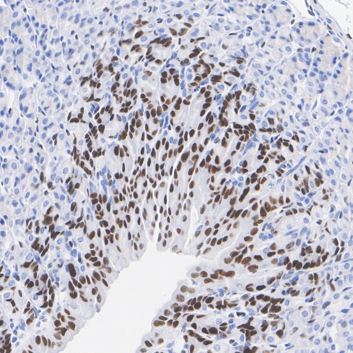
Fig6: Species: Mouse, Tissue: Stomach, Sample: Paraffin-embedded section.Application
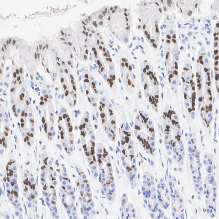
Fig7: Species: Rat, Tissue: Stomach, Sample: Paraffin-embedded section.| Product Name | KLF4 Recombinant Rabbit Monoclonal Antibody |
|---|---|
| Antibody Type | Primary Antibodies |
| Product description | The Kruppel-type zinc finger transcription factors comprise a conserved family of DNA binding proteins that are important in developmental regulation. The Kruppel zinc finger transcription factor was initially identified in Drosophila as a segmentation gene. Kruppel-like factors that have been characterized in mammals include EKLF, LKLF and GKLF (6-8). EKLF is expressed principally in erythroid tissues, and LKLF expression is limited to the lung. GKLF is found predominantly in gut and has been shown to be expressed during growth arres. |
| Immunogen | Synthetic peptide within human KLF4 aa 320-360. |
| Clonality | monoclonal |
|---|---|
| Isotype | IgG |
| Host Species | Recombinant rabbit |
| Tested Applications | ICC/IFIHCWB |
| WB:1:500-1:2000 ICC:1:50-1:200 IHC:1:50-1:200 |
|
| Species Reactivity | HumanMouse |
| Concentration | 1mg/ml |
| Purification | Affinity purified |
| Gene Symbol | KLF4 |
|---|---|
| Gene Synonyms | EZF GKLF |
| Gene Full Name | KLF transcription factor 4 |
| Alternative Names | Endothelial Kruppel like zinc finger protein antibody Epithelial zinc finger protein EZF antibody EZF antibody GKLF antibody gut Kruppel-like factor antibody Gut-enriched krueppel-like factor antibody KLF antibody KLF4 antibody KLF4_HUMAN antibody Krueppel-like factor 4 antibody Kruppel like factor 4 (Epithelial zinc finger protein EZF) (Gut enriched Krueppel like factor) antibody Kruppel like factor 4 (gut) antibody |
| Molecular Weight(MW) | 55kDa |
| Cellular Localization | Nucleus. |
Application
Fig1: Western blot analysis of KLF4 on different lysates with Rabbit anti-KLF4 antibody at 1/2,000 dilution. Lane 1: 293 cell lysate, Lane 2: NCCIT cell lysate, Lysates/proteins at 10 µg/Lane. Exposure time: 1 minute 30 seconds; 10% SDS-PAGE gel. Proteins were transferred to a PVDF membrane and blocked with 5% NFDM/TBST for 1 hour at room temperature. The primary antibody at 1/2,000 dilution was used in 5% NFDM/TBST at room temperature for 2 hours. Goat Anti-Rabbit IgG - HRP Secondary Antibody at 1:100,000 dilution was used for 1 hour at room temperature.
Application
Fig2: ICC staining KLF4 in Hela cells (red). The nuclear counter stain is DAPI (blue). Cells were fixed in paraformaldehyde, permeabilised with 0.25% Triton X100/PBS.
Application
Fig3: ICC staining KLF4 in PC-3M cells (red). The nuclear counter stain is DAPI (blue). Cells were fixed in paraformaldehyde, permeabilised with 0.25% Triton X100/PBS.
Application
Fig4: ICC staining KLF4 in 293T cells (red). The nuclear counter stain is DAPI (blue). Cells were fixed in paraformaldehyde, permeabilised with 0.25% Triton X100/PBS.
Application
Fig5: Species: Human, Tissue: Stomach, Sample: Paraffin-embedded section.
Application
Fig6: Species: Mouse, Tissue: Stomach, Sample: Paraffin-embedded section.
Application
Fig7: Species: Rat, Tissue: Stomach, Sample: Paraffin-embedded section.| Positive Control | 293, NCCIT, 293T, PC-3M, Hela, human liver tissue, mouse liver tissue, human gastric carcinoma tissue, mouse colon tissue, mouse stomach tissue. |
|---|---|
| Application Notes | WB:1:500-1:2000 ICC:1:50-1:200 IHC:1:50-1:200 |
| Form | Liquid |
|---|---|
| Storage Instructions | Store at +4℃ after thawing. Aliquot store at -20℃ or -80℃. Avoid repeated freeze / thaw cycles. |
| Storage Buffer | 1*TBS (pH7.4), 1%BSA, 40%Glycerol. Preservative: 0.05% Sodium Azide. |


