WB
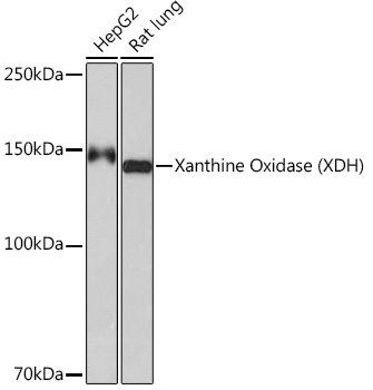
Western blot analysis of various lysates using Xanthine Oxidase (XDH) Rabbit mAb at 1:1000 dilution. Secondary antibody: HRP-conjugated Goat anti-Rabbit IgG (H+L) at 1:10000 dilution. Lysates/proteins: 25μg per lane. Blocking buffer: 3% nonfat dry milk in TBST. Detection: ECL Basic Kit. Exposure time: 60s.IHC
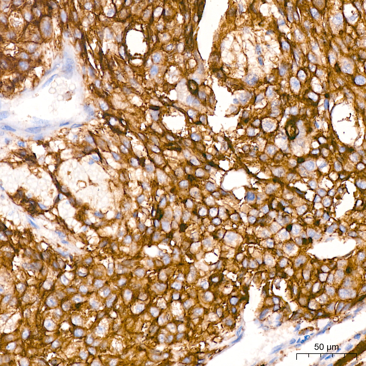
Immunohistochemistry analysis of paraffin embedded Human liver cancer using Xanthine Oxidase (XDH) Rabbit mAb at dilution of 1:200 (40x lens). High pressure antigen retrieval performed with 0.01M Citrate buffer (pH 6.0) prior to IHC staining.IHC
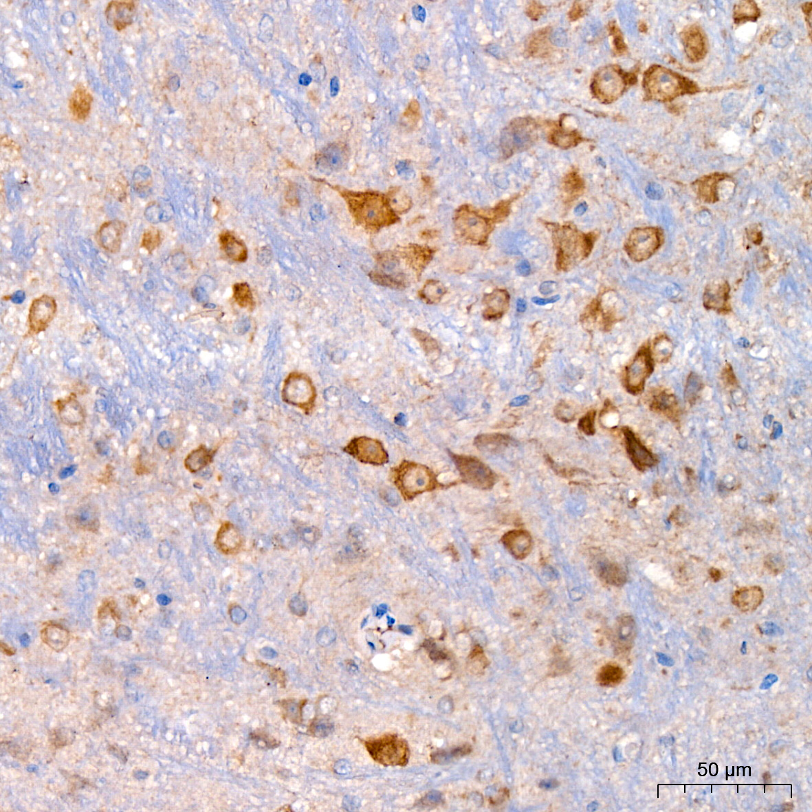
Immunohistochemistry analysis of paraffin embedded Mouse brain using Xanthine Oxidase (XDH) Rabbit mAb at dilution of 1:200 (40x lens). High pressure antigen retrieval performed with 0.01M Citrate buffer (pH 6.0) prior to IHC staining.ICC/IF
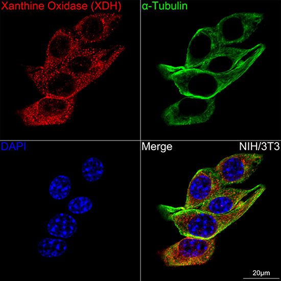
Confocal imaging of NIH/3T3 cells using Xanthine Oxidase (XDH) Rabbit mAb (dilution 1:200) followed by a further incubation with Cy3 Goat Anti-Rabbit IgG (H+L) (dilution 1:500) (Red). The cells were counterstained with α-Tubulin Mouse mAb (dilution 1:400) followed by incubation with Omnimabs® 488 Goat Anti-Mouse IgG(H&L) Ab (dilution 1:500) (Green). DAPI was used for nuclear staining (Blue). Objective: 100x.IF-P
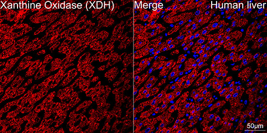
Confocal imaging of paraffin-embedded Human liver using Xanthine Oxidase (XDH) Rabbit mAb (dilution 1:200) followed by a further incubation with Cy3 Goat Anti Rabbit IgG (H+L) (dilution 1:500) (Red). DAPI was used for nuclear staining (Blue). Objective: 40x. Perform high pressure antigen retrieval with 0.01 M citrate buffer (pH 6.0) prior to IF staining.| Product Name | Xanthine Oxidase (XDH) Rabbit mAb |
|---|---|
| Antibody Type | Primary Antibodies |
| Product description | The process of metabolizing purines to a common molecule known as xanthine is an essential process for the proper shuttling of uric acid. Xanthine oxidase is a flavoprotein enzyme that coordinates molybdenum and utilizes NAD+ as an electron acceptor to catalyze the oxidation of hypoxanthine to xanthine and then to uric acid. The predominant form of this enzyme is xanthine dehydrogenase, which is a homodimer that can be converted to xanthine oxidase by sulfhydryl oxidation or proteolytic modification. Xanthine oxidase is present in species ranging from bacteria to human and is ubiquitously expressed in mammalian tissues. In the oxidase form, this enzyme is coupled to the generation of free radicals. Individuals showing marked elevation of serum xanthine oxidase is suggestive of chronic liver disease and cholestasis, which is a condition defined by hepatic obstruction. Hepatic obstruction causes bile salts, the bile pigment bilirubin, and fats to accumulate in the blood stream instead of being eliminated normally. The clinical consequences of defects in xanthine oxidase range from mild to severe and even contribute to fatal disorders. |
| Immunogen | Recombinant fusion protein containing a sequence corresponding to amino acids 202-293 of human Xanthine Oxidase (XDH) (P47989). |
| Clonality | Monoclonal |
|---|---|
| Isotype | IgG |
| Host Species | Recombinant rabbit |
| Tested Applications | ICC/IFIF-PIHCWB |
| WB:1:1000-1:2000 IHC:1:200-1:2000 ICC/IF:1:200-1:400 IF-P:1:200-1:400 |
|
| Species Reactivity | HumanMouseRat |
| Concentration | 1mg/ml |
| Purification | Affinity purified |
| Gene Symbol | XDH |
|---|---|
| Gene Synonyms | XO XOR XAN1 |
| Gene Full Name | xanthine dehydrogenase |
| Molecular Weight(MW) | 146kDa(Observed MW 150kDa) |
| Cellular Localization | Cytoplasm, Peroxisome, Secreted. |
WB
Western blot analysis of various lysates using Xanthine Oxidase (XDH) Rabbit mAb at 1:1000 dilution. Secondary antibody: HRP-conjugated Goat anti-Rabbit IgG (H+L) at 1:10000 dilution. Lysates/proteins: 25μg per lane. Blocking buffer: 3% nonfat dry milk in TBST. Detection: ECL Basic Kit. Exposure time: 60s.
IHC
Immunohistochemistry analysis of paraffin embedded Human liver cancer using Xanthine Oxidase (XDH) Rabbit mAb at dilution of 1:200 (40x lens). High pressure antigen retrieval performed with 0.01M Citrate buffer (pH 6.0) prior to IHC staining.
IHC
Immunohistochemistry analysis of paraffin embedded Mouse brain using Xanthine Oxidase (XDH) Rabbit mAb at dilution of 1:200 (40x lens). High pressure antigen retrieval performed with 0.01M Citrate buffer (pH 6.0) prior to IHC staining.
ICC/IF
Confocal imaging of NIH/3T3 cells using Xanthine Oxidase (XDH) Rabbit mAb (dilution 1:200) followed by a further incubation with Cy3 Goat Anti-Rabbit IgG (H+L) (dilution 1:500) (Red). The cells were counterstained with α-Tubulin Mouse mAb (dilution 1:400) followed by incubation with Omnimabs® 488 Goat Anti-Mouse IgG(H&L) Ab (dilution 1:500) (Green). DAPI was used for nuclear staining (Blue). Objective: 100x.
IF-P
Confocal imaging of paraffin-embedded Human liver using Xanthine Oxidase (XDH) Rabbit mAb (dilution 1:200) followed by a further incubation with Cy3 Goat Anti Rabbit IgG (H+L) (dilution 1:500) (Red). DAPI was used for nuclear staining (Blue). Objective: 40x. Perform high pressure antigen retrieval with 0.01 M citrate buffer (pH 6.0) prior to IF staining.| Application Notes | WB:1:1000-1:2000 IHC:1:200-1:2000 ICC/IF:1:200-1:400 IF-P:1:200-1:400 |
|---|
| Form | Liquid |
|---|---|
| Storage Instructions | Store at -20℃. Avoid freeze / thaw cycles. |
| Storage Buffer | PBS with 0.02% sodium azide, 0.05% BSA, 50% glycerol, pH7.3. |


