Application
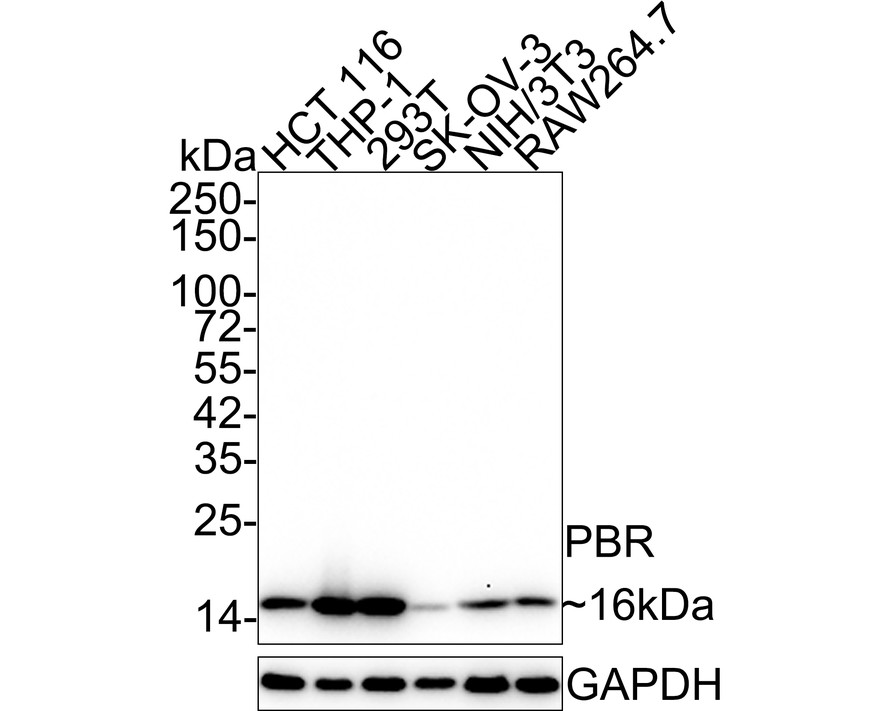
Fig1: Western blot analysis of PBR on different lysates with Rabbit anti-PBR antibody at 1/2,000 dilution. Lane 1: HCT 116 cell lysate, Lane 2: THP-1 cell lysate, Lane 3: 293T cell lysate, Lane 4: SK-OV-3 cell lysate, Lane 5: NIH/3T3 cell lysate, Lane 6: RAW264.7 cell lysate, Lysates/proteins at 15 µg/Lane. Exposure time: 1 minute 4 seconds; 4-20% SDS-PAGE gel. Proteins were transferred to a PVDF membrane and blocked with 5% NFDM/TBST for 1 hour at room temperature. The primary antibody at 1/2,000 dilution was used in 5% NFDM/TBST at 4℃ overnight. Goat Anti-Rabbit IgG - HRP Secondary Antibody at 1/50,000 dilution was used for 1 hour at room temperature.Application
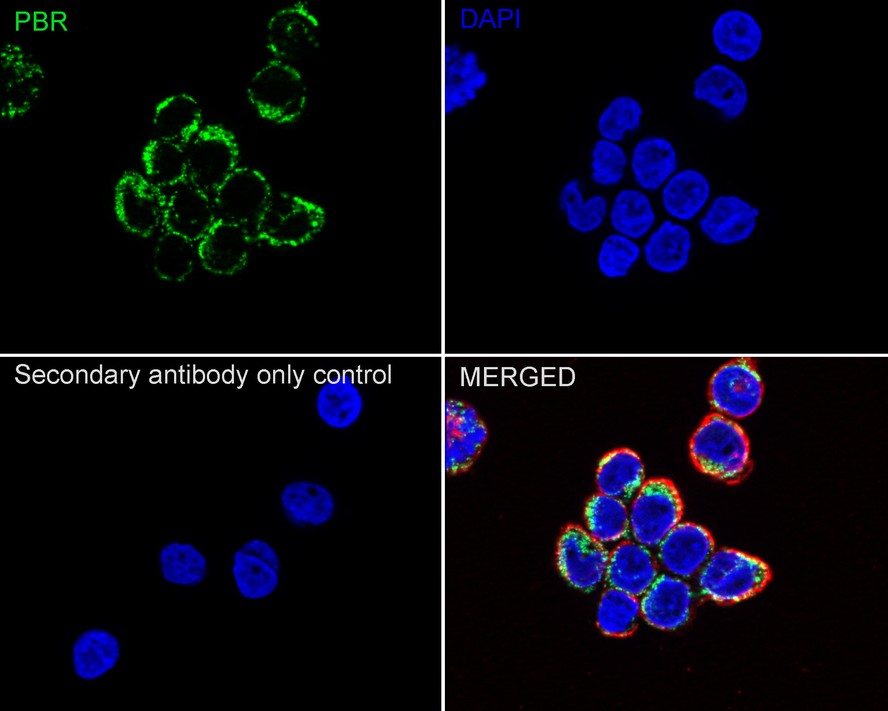
Fig2: Immunocytochemistry analysis of THP-1 cells labeling PBR with Rabbit anti-PBR antibody at 1/1,000 dilution. Cells were fixed in 4% paraformaldehyde for 20 minutes at room temperature, permeabilized with 0.1% Triton X-100 in PBS for 5 minutes at room temperature, then blocked with 1% BSA in 10% negative goat serum for 1 hour at room temperature. Cells were then incubated with Rabbit anti-PBR antibody at 1/1,000 dilution in 1% BSA in PBST overnight at 4 ℃. Goat Anti-Rabbit IgG H&L (iFluor™ 488) was used as the secondary antibody at 1/1,000 dilution. PBS instead of the primary antibody was used as the secondary antibody only control. Nuclear DNA was labelled in blue with DAPI. Beta tubulin (red) was stained at 1/100 dilution overnight at +4℃. Goat Anti-Mouse IgG H&L (iFluor™ 594) was used as the secondary antibody at 1/1,000 dilution.Application
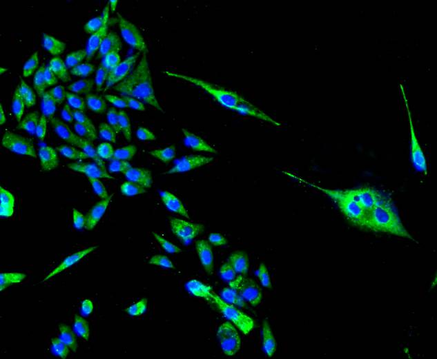
Fig3: ICC staining PBR in PC-3M cells (green). The nuclear counter stain is DAPI (blue). Cells were fixed in paraformaldehyde, permeabilised with 0.25% Triton X100/PBS.Application
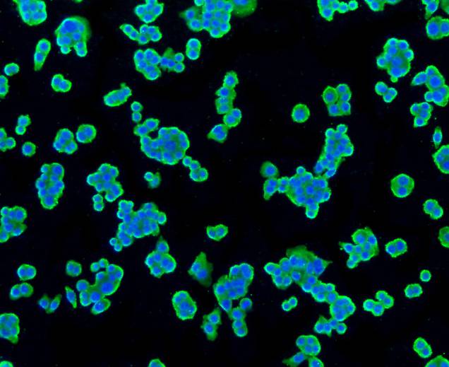
Fig4: ICC staining PBR in SW480 cells (green). Cells were fixed in paraformaldehyde, permeabilised with 0.25% Triton X100/PBS.Application
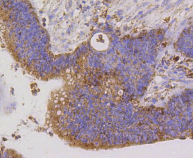
Fig5: Immunohistochemical analysis of paraffin-embedded human colon cancer tissue using anti-PBR antibody. Counter stained with hematoxylin.Application
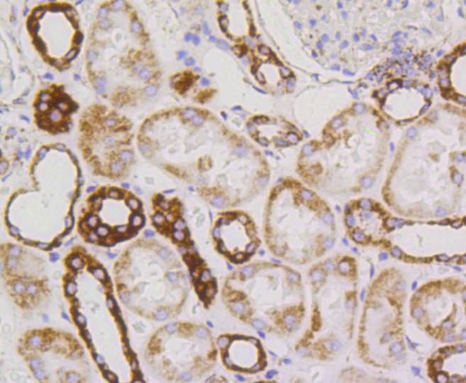
Fig6: Immunohistochemical analysis of paraffin-embedded human kidney tissue using anti-PBR antibody. Counter stained with hematoxylin.Application
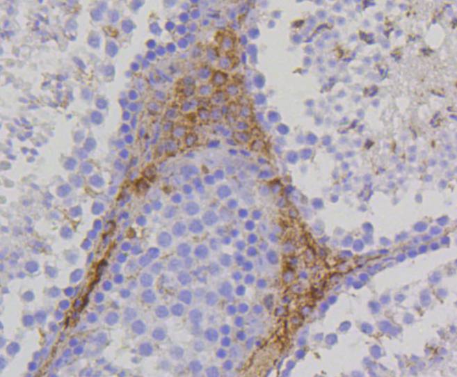
Fig7: Immunohistochemical analysis of paraffin-embedded mouse testis tissue using anti-PBR antibody. Counter stained with hematoxylin.Application
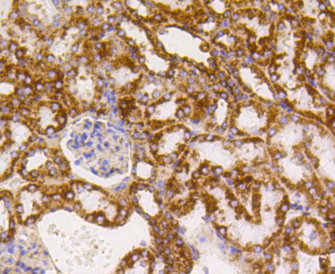
Fig8: Immunohistochemical analysis of paraffin-embedded mouse kidney tissue using anti-PBR antibody. Counter stained with hematoxylin.Application
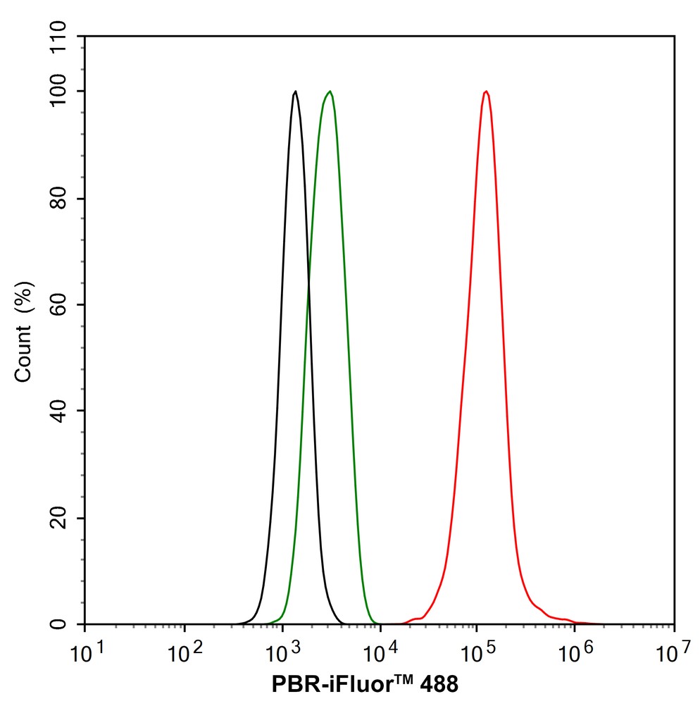
Fig9: Flow cytometric analysis of THP-1 cells labeling PBR. Cells were fixed and permeabilized. Then stained with the primary antibody (1/1,000) (red) compared with Rabbit IgG Isotype Control (green). After incubation of the primary antibody at +4℃ for an hour, the cells were stained with a iFluor™ 488 conjugate-Goat anti-Rabbit IgG Secondary antibody at 1/1,000 dilution for 30 minutes at +4℃. Unlabelled sample was used as a control (cells without incubation with primary antibody; black).Application
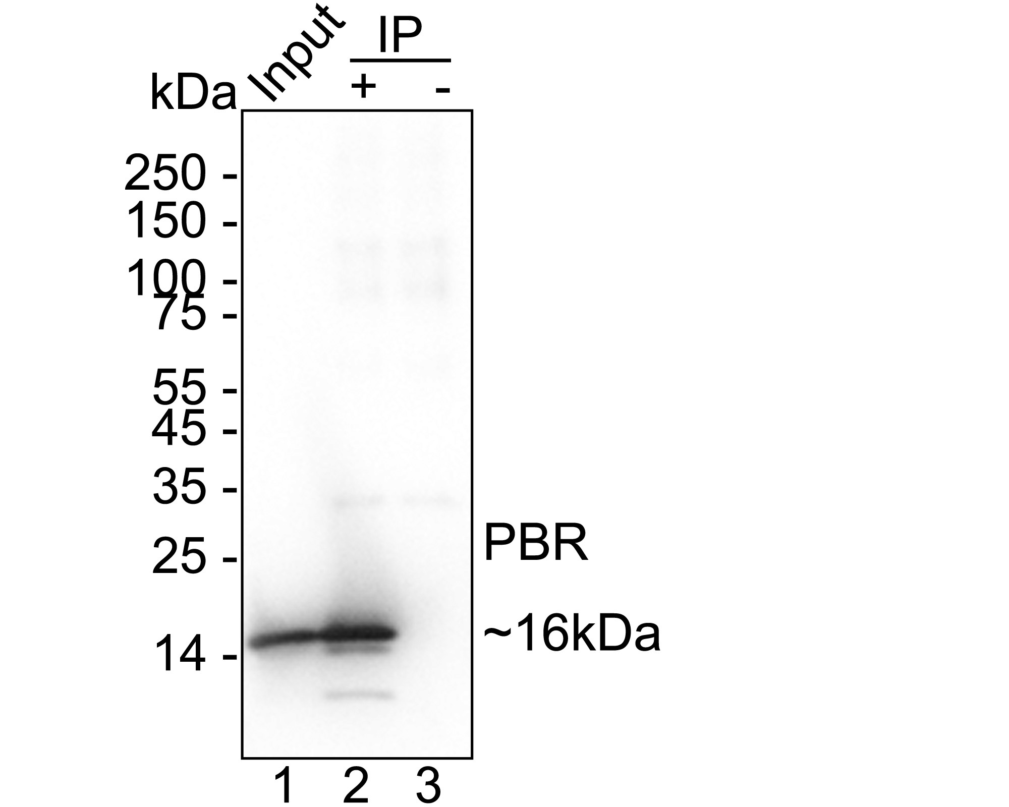
Fig10:PBR was immunoprecipitated from 0.2 mg THP-1 cell lysate with Rabbit anti-PBR antibody at 2 µg/25 µl agarose. Western blot was performed from the immunoprecipitate using Rabbit anti-PBR antibody at 1/1,000 dilution. Anti-Rabbit IgG for IP Nano-secondary antibody at 1/5,000 dilution was used for 1 hour at room temperature. Lane 1: THP-1 cell lysate (input), Lane 2: Rabbit anti-PBR antibody IP in THP-1 cell lysate, Lane 3: Rabbit IgG instead of Rabbit anti-PBR antibody in THP-1 cell lysate, Blocking/Dilution buffer: 5% NFDM/TBST. Exposure time: 1 seconds.| Product Name | PBR Recombinant Rabbit Monoclonal Antibody |
|---|---|
| Antibody Type | Primary Antibodies |
| Product description | Mitochondrial peripheral-type benzodiazepine receptor (PBR) is an indispensable element of the steroidogenic machinery, where it mediates the delivery of cholesterol to the inner mitochondrial side chain cleavage cytochrome P-450 upon ligand activation. PBR is composed of three subunits, an isoquinoline binding site, a voltage-dependent anion channel and an adenine nucleotide carrier. PBR is genetically conserved from bacteria to humans and in humans is widely expressed in peripheral organs, whereas in the brain, it is sparse and located mainly in glial cells. Peroxisome proliferator perfluordecanoic acid (PFDA) inhibits the Leydig cell steroidogenesis by affecting PBR mRNA stability, thus inhibiting PBR expression, cholesterol transport into the mitochondria and subsequent steroid formation. A cytoplasmic protein, PRAX-1 ( peripheral benzodiazepine receptor-associated protein 1), is found to specifically interact with PBR. The polypeptide diazepam binding inhibitor is an endogenous PBR ligand. PBR also binds Ro 5-4864 (4’-chlorodiazepam) and PK 11185 (an isoquinoline carboxamide derivative), but not clonazepam, and PBR regulates the cholesterol transport that results in decreased circulating corticosterone levels. |
| Immunogen | Synthetic peptide within Human PBR aa 120-169 / 169. |
| Clonality | monoclonal |
|---|---|
| Isotype | IgG |
| Host Species | Recombinant rabbit |
| Tested Applications | FCICC/IFIHCIPWB |
| WB:1:2000 ICC:1:1000 IHC:1:400-1:1000 FC:1:1000 IP:1-2μg/sample |
|
| Species Reactivity | HumanMouseRat |
| Concentration | 1mg/ml |
| Purification | Protein A |
| Gene Symbol | TSPO |
|---|---|
| Gene Synonyms | DBI IBP MBR PBR PBS BPBS BZRP PKBS PTBR mDRC pk18 TSPO1 |
| Gene Full Name | translocator protein |
| Gene Summary | Present mainly in the mitochondrial compartment of peripheral tissues, the protein encoded by this gene interacts with some benzodiazepines and has different affinities than its endogenous counterpart. The protein is a key factor in the flow of cholesterol into mitochondria to permit the initiation of steroid hormone synthesis. Alternatively spliced transcript variants have been reported; one of the variants lacks an internal exon and is considered non-coding, and the other variants encode the same protein. [provided by RefSeq, Feb 2012] |
| Alternative Names | Benzodiazapine receptor (peripheral) antibody Benzodiazepine peripheral binding site antibody BPBS antibody BZRP antibody DBI antibody IBP antibody Isoquinoline carboxamide-binding protein antibody MBR antibody mDRC antibody Mitochondrial benzodiazepine receptor antibody PBR antibody PBS antibody Peripheral benzodiazepine receptor antibody Peripheral benzodiazepine receptor-related protein antibody Peripheral type benzodiazepine receptor antibody Peripheral-type benzodiazepine receptor antibody pk18 antibody PKBS antibody PTBR antibody Ptbzr antibody PTBZR02 antibody RATPTBZR02 antibody translocator protein (18kDa) antibody Translocator protein antibody Tspo antibody Tspo1 antibody TSPOA_HUMAN antibody |
| Molecular Weight(MW) | 19kDa(Observed band size: 16 kDa) |
| Cellular Localization | Mitochondrion membrane |
Application
Fig1: Western blot analysis of PBR on different lysates with Rabbit anti-PBR antibody at 1/2,000 dilution. Lane 1: HCT 116 cell lysate, Lane 2: THP-1 cell lysate, Lane 3: 293T cell lysate, Lane 4: SK-OV-3 cell lysate, Lane 5: NIH/3T3 cell lysate, Lane 6: RAW264.7 cell lysate, Lysates/proteins at 15 µg/Lane. Exposure time: 1 minute 4 seconds; 4-20% SDS-PAGE gel. Proteins were transferred to a PVDF membrane and blocked with 5% NFDM/TBST for 1 hour at room temperature. The primary antibody at 1/2,000 dilution was used in 5% NFDM/TBST at 4℃ overnight. Goat Anti-Rabbit IgG - HRP Secondary Antibody at 1/50,000 dilution was used for 1 hour at room temperature.
Application
Fig2: Immunocytochemistry analysis of THP-1 cells labeling PBR with Rabbit anti-PBR antibody at 1/1,000 dilution. Cells were fixed in 4% paraformaldehyde for 20 minutes at room temperature, permeabilized with 0.1% Triton X-100 in PBS for 5 minutes at room temperature, then blocked with 1% BSA in 10% negative goat serum for 1 hour at room temperature. Cells were then incubated with Rabbit anti-PBR antibody at 1/1,000 dilution in 1% BSA in PBST overnight at 4 ℃. Goat Anti-Rabbit IgG H&L (iFluor™ 488) was used as the secondary antibody at 1/1,000 dilution. PBS instead of the primary antibody was used as the secondary antibody only control. Nuclear DNA was labelled in blue with DAPI. Beta tubulin (red) was stained at 1/100 dilution overnight at +4℃. Goat Anti-Mouse IgG H&L (iFluor™ 594) was used as the secondary antibody at 1/1,000 dilution.
Application
Fig3: ICC staining PBR in PC-3M cells (green). The nuclear counter stain is DAPI (blue). Cells were fixed in paraformaldehyde, permeabilised with 0.25% Triton X100/PBS.
Application
Fig4: ICC staining PBR in SW480 cells (green). Cells were fixed in paraformaldehyde, permeabilised with 0.25% Triton X100/PBS.
Application
Fig5: Immunohistochemical analysis of paraffin-embedded human colon cancer tissue using anti-PBR antibody. Counter stained with hematoxylin.
Application
Fig6: Immunohistochemical analysis of paraffin-embedded human kidney tissue using anti-PBR antibody. Counter stained with hematoxylin.
Application
Fig7: Immunohistochemical analysis of paraffin-embedded mouse testis tissue using anti-PBR antibody. Counter stained with hematoxylin.
Application
Fig8: Immunohistochemical analysis of paraffin-embedded mouse kidney tissue using anti-PBR antibody. Counter stained with hematoxylin.
Application
Fig9: Flow cytometric analysis of THP-1 cells labeling PBR. Cells were fixed and permeabilized. Then stained with the primary antibody (1/1,000) (red) compared with Rabbit IgG Isotype Control (green). After incubation of the primary antibody at +4℃ for an hour, the cells were stained with a iFluor™ 488 conjugate-Goat anti-Rabbit IgG Secondary antibody at 1/1,000 dilution for 30 minutes at +4℃. Unlabelled sample was used as a control (cells without incubation with primary antibody; black).
Application
Fig10:PBR was immunoprecipitated from 0.2 mg THP-1 cell lysate with Rabbit anti-PBR antibody at 2 µg/25 µl agarose. Western blot was performed from the immunoprecipitate using Rabbit anti-PBR antibody at 1/1,000 dilution. Anti-Rabbit IgG for IP Nano-secondary antibody at 1/5,000 dilution was used for 1 hour at room temperature. Lane 1: THP-1 cell lysate (input), Lane 2: Rabbit anti-PBR antibody IP in THP-1 cell lysate, Lane 3: Rabbit IgG instead of Rabbit anti-PBR antibody in THP-1 cell lysate, Blocking/Dilution buffer: 5% NFDM/TBST. Exposure time: 1 seconds.| Positive Control | PC-3M, MCF-7, Hela, HepG2, SW480, NIH/3T3, mouse kidney tissue, human colon cancer tissue, human kidney tissue, mouse testis tissue. |
|---|---|
| Application Notes | WB:1:2000 ICC:1:1000 IHC:1:400-1:1000 FC:1:1000 IP:1-2μg/sample |
| Form | Liquid |
|---|---|
| Storage Instructions | Store at +4℃ after thawing. Aliquot store at -20℃ or -80℃. Avoid repeated freeze / thaw cycles. |
| Storage Buffer | 1*TBS (pH7.4), 1%BSA, 40%Glycerol. Preservative: 0.05% Sodium Azide. |


