WB
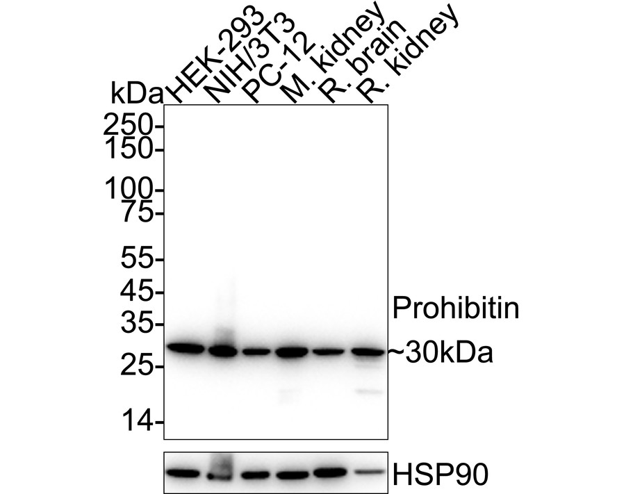
Western blot analysis of Prohibitin on different lysates with Rabbit anti-Prohibitin antibody at 1/1,000 dilution. Lane 1: HEK-293 cell lysate (20 µg/Lane), Lane 2: NIH/3T3 cell lysate (20 µg/Lane), Lane 3: PC-12 cell lysate (20 µg/Lane), Lane 4: Mouse kidney tissue lysate (40 µg/Lane), Lane 5: Rat brain tissue lysate (40 µg/Lane), Lane 6: Rat kidney tissue lysate (40 µg/Lane), Exposure time: 10 seconds; 4-20% SDS-PAGE gel. Proteins were transferred to a PVDF membrane and blocked with 5% NFDM/TBST for 1 hour at room temperature. The primary antibody at 1/1,000 dilution was used in 5% NFDM/TBST at 4℃ overnight. Goat Anti-Rabbit IgG - HRP Secondary Antibody at 1/50,000 dilution was used for 1 hour at room temperature.IHC
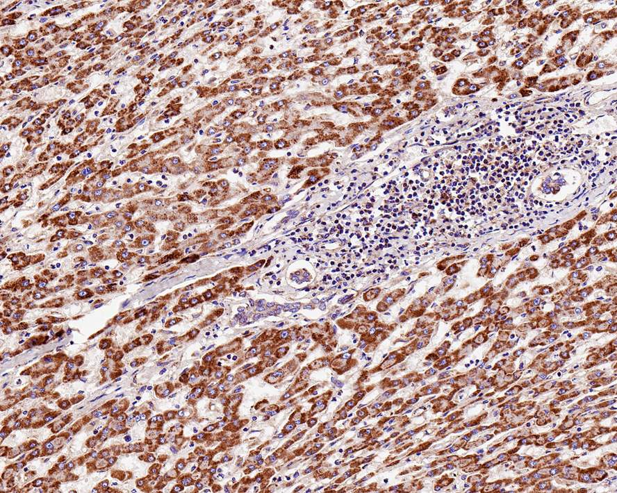
Immunohistochemical analysis of paraffin-embedded human liver tissue with Rabbit anti-Prohibitin antibody at 1/200 dilution. The section was pre-treated using heat mediated antigen retrieval with Tris-EDTA buffer (pH 9.0) for 20 minutes. The tissues were blocked in 1% BSA for 20 minutes at room temperature, washed with ddH2O and PBS, and then probed with the primary antibody at 1/200 dilution for 1 hour at room temperature. The detection was performed using an HRP conjugated compact polymer system. DAB was used as the chromogen. Tissues were counterstained with hematoxylin and mounted with DPX.IHC
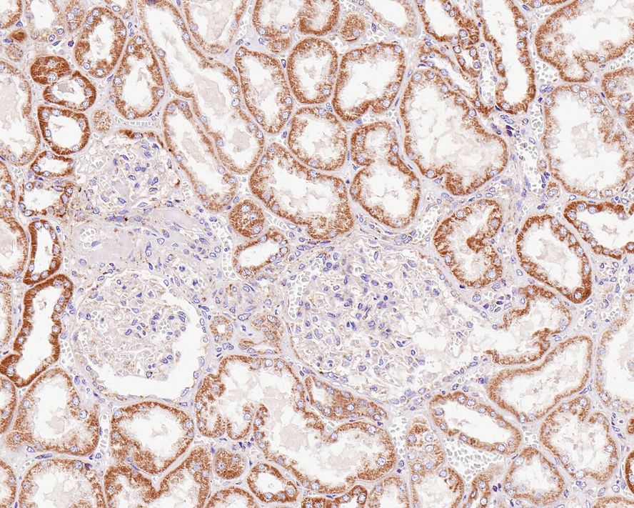
Immunohistochemical analysis of paraffin-embedded human kidney tissue with Rabbit anti-Prohibitin antibody at 1/1,000 dilution. The section was pre-treated using heat mediated antigen retrieval with Tris-EDTA buffer (pH 9.0) for 20 minutes. The tissues were blocked in 1% BSA for 20 minutes at room temperature, washed with ddH2O and PBS, and then probed with the primary antibody at 1/1,000 dilution for 1 hour at room temperature. The detection was performed using an HRP conjugated compact polymer system. DAB was used as the chromogen. Tissues were counterstained with hematoxylin and mounted with DPX.IHC
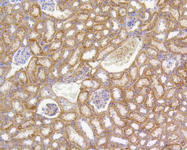
Immunohistochemical analysis of paraffin-embedded mouse kidney tissue using anti-Prohibitin antibody. The section was pre-treated using heat mediated antigen retrieval with Tris-EDTA buffer (pH 8.0-8.4) for 20 minutes.The tissues were blocked in 5% BSA for 30 minutes at room temperature, washed with ddH2O and PBS, and then probed with the primary antibody (1/200) for 30 minutes at room temperature. The detection was performed using an HRP conjugated compact polymer system. DAB was used as the chromogen. Tissues were counterstained with hematoxylin and mounted with DPX.IHC
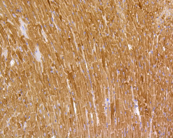
Immunohistochemical analysis of paraffin-embedded mouse heart tissue using anti-Prohibitin antibody. The section was pre-treated using heat mediated antigen retrieval with Tris-EDTA buffer (pH 8.0-8.4) for 20 minutes.The tissues were blocked in 5% BSA for 30 minutes at room temperature, washed with ddH2O and PBS, and then probed with the primary antibody (1/200) for 30 minutes at room temperature. The detection was performed using an HRP conjugated compact polymer system. DAB was used as the chromogen. Tissues were counterstained with hematoxylin and mounted with DPX.ICC/IF
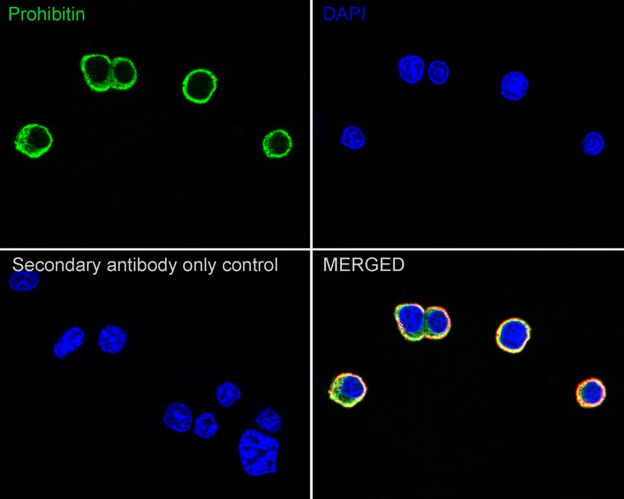
Immunocytochemistry analysis of PC-12 cells labeling Prohibitin with Rabbit anti-Prohibitin antibody at 1/1,000 dilution. Cells were fixed in 4% paraformaldehyde for 15 minutes at room temperature, permeabilized with 0.1% Triton X-100 in PBS for 15 minutes at room temperature, then blocked with 1% BSA in 10% negative goat serum for 1 hour at room temperature. Cells were then incubated with Rabbit anti-Prohibitin antibody at 1/1,000 dilution in 1% BSA in PBST overnight at 4 ℃. Goat Anti-Rabbit IgG H&L (488) was used as the secondary antibody at 1/1,000 dilution. PBS instead of the primary antibody was used as the secondary antibody only control. Nuclear DNA was labelled in blue with DAPI. Beta tubulin (red) was stained at 1/100 dilution overnight at +4℃. Goat Anti-Mouse IgG H&L (594) was used as the secondary antibody at 1/1,000 dilution.FC
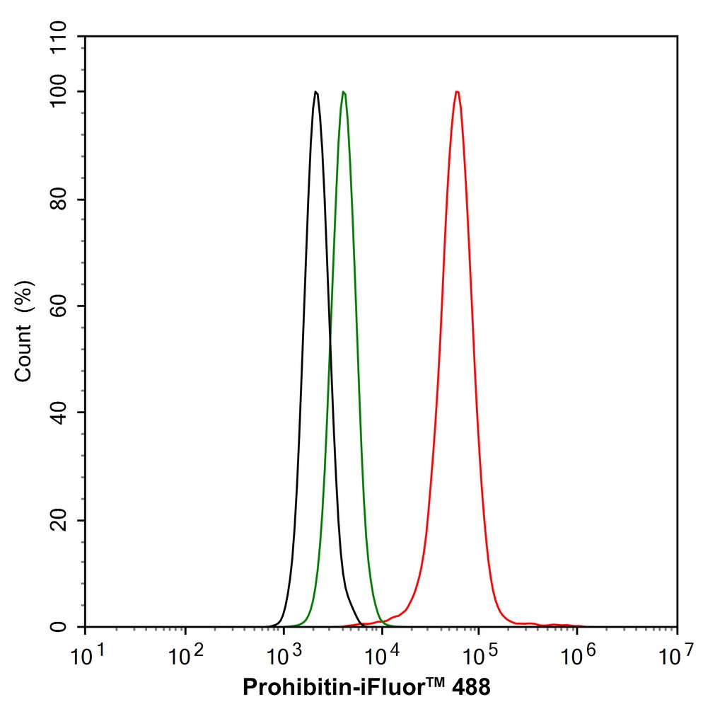
Flow cytometric analysis of HepG2 cells labeling Prohibitin. Cells were fixed and permeabilized. Then stained with the primary antibody (1μg/mL) (red) compared with Rabbit IgG Isotype Control (green). After incubation of the primary antibody at +4℃ for an hour, the cells were stained with a 488 conjugate-Goat anti-Rabbit IgG Secondary antibody at 1/1,000 dilution for 30 minutes at +4℃. Unlabelled sample was used as a control (cells without incubation with primary antibody; black).IP
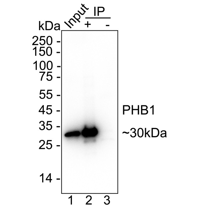
Prohibitin was immunoprecipitated from 0.2 mg HEK-293 cell lysate with Rabbit anti-Prohibitin antibody at 2 µg/10 µl beads. Western blot was performed from the immunoprecipitate using Rabbit anti-Prohibitin antibody at 1/2,000 dilution. HRP Conjugated Anti-Rabbit IgG for IP Nano-secondary antibody at 1/5,000 dilution was used for 1 hour at room temperature. Lane 1: HEK-293 cell lysate (input), Lane 2:Rabbit anti-Prohibitin antibody IP in HEK-293 cell lysate, Lane 3: Rabbit IgG instead of Rabbit anti-Prohibitin antibody in HEK-293 cell lysate, Blocking/Dilution buffer: primary antibody dilution, Exposure time: 3 seconds.| Product Name | Prohibitin Recombinant Rabbit Monoclonal Antibody |
|---|---|
| Antibody Type | Primary Antibodies |
| Product description | Prohibitin is an evolutionarily conserved protein that has antiproliferative activity. The gene encoding human prohibitin maps to chromosome 17q21 and is ubiquitously expressed. Prohibitin is a post-synthetically modified protein that is localized in the inner membrane of mitochondria, where it regulates the cell cycle by blocking the transition between the G1 and S phases, and on the plasma membrane of B cells, where it mediates B cell maturation. Prohibitin mRNA and protein levels are high in G1, decline during the S phase, rise again in G2 and decline in M phase, which suggests that prohibitin controls the cell cycle by using both transcriptional and posttranslational mechanisms. Prohibitin is also a potential tumor suppressor protein that binds to retinoblastoma (Rb) and subsequently inhibits the activity of E2F family members in response to specific signaling cascades. Prohibitin 2 is a repressor of estrogen receptor activity, and is required for somatic and germline differentiation in the larval gonad during embryonic development. Mutations in the Prohibitin genes are correlated with breast cancer development and/or progressionin more than 80% of the cell lines analyzed. |
| Immunogen | Synthetic peptide within Human Prohibitin aa 147-196 / 272. |
| Clonality | Monoclonal |
|---|---|
| Isotype | IgG |
| Host Species | Recombinant rabbit |
| Tested Applications | FCICC/IFIHCIPWB |
| WB:1:1000 IHC:1:200-1:1000 ICC/IF:1:100-1:1000 FC:1:1000 IP:1-2μg/sample |
|
| Species Reactivity | HumanMouseRat |
| Concentration | 1mg/ml |
| Purification | Protein A |
| Gene Symbol | PHB1 |
|---|---|
| Gene Synonyms | PHB BAP32 HEL-215 HEL-S-54e |
| Gene Full Name | prohibitin 1 |
| Molecular Weight(MW) | 30 kDa |
| Cellular Localization | Cell membrane, Cytoplasm, Membrane, Mitochondrion, Mitochondrion inner membrane, Nucleus. |
WB
Western blot analysis of Prohibitin on different lysates with Rabbit anti-Prohibitin antibody at 1/1,000 dilution. Lane 1: HEK-293 cell lysate (20 µg/Lane), Lane 2: NIH/3T3 cell lysate (20 µg/Lane), Lane 3: PC-12 cell lysate (20 µg/Lane), Lane 4: Mouse kidney tissue lysate (40 µg/Lane), Lane 5: Rat brain tissue lysate (40 µg/Lane), Lane 6: Rat kidney tissue lysate (40 µg/Lane), Exposure time: 10 seconds; 4-20% SDS-PAGE gel. Proteins were transferred to a PVDF membrane and blocked with 5% NFDM/TBST for 1 hour at room temperature. The primary antibody at 1/1,000 dilution was used in 5% NFDM/TBST at 4℃ overnight. Goat Anti-Rabbit IgG - HRP Secondary Antibody at 1/50,000 dilution was used for 1 hour at room temperature.
IHC
Immunohistochemical analysis of paraffin-embedded human liver tissue with Rabbit anti-Prohibitin antibody at 1/200 dilution. The section was pre-treated using heat mediated antigen retrieval with Tris-EDTA buffer (pH 9.0) for 20 minutes. The tissues were blocked in 1% BSA for 20 minutes at room temperature, washed with ddH2O and PBS, and then probed with the primary antibody at 1/200 dilution for 1 hour at room temperature. The detection was performed using an HRP conjugated compact polymer system. DAB was used as the chromogen. Tissues were counterstained with hematoxylin and mounted with DPX.
IHC
Immunohistochemical analysis of paraffin-embedded human kidney tissue with Rabbit anti-Prohibitin antibody at 1/1,000 dilution. The section was pre-treated using heat mediated antigen retrieval with Tris-EDTA buffer (pH 9.0) for 20 minutes. The tissues were blocked in 1% BSA for 20 minutes at room temperature, washed with ddH2O and PBS, and then probed with the primary antibody at 1/1,000 dilution for 1 hour at room temperature. The detection was performed using an HRP conjugated compact polymer system. DAB was used as the chromogen. Tissues were counterstained with hematoxylin and mounted with DPX.
IHC
Immunohistochemical analysis of paraffin-embedded mouse kidney tissue using anti-Prohibitin antibody. The section was pre-treated using heat mediated antigen retrieval with Tris-EDTA buffer (pH 8.0-8.4) for 20 minutes.The tissues were blocked in 5% BSA for 30 minutes at room temperature, washed with ddH2O and PBS, and then probed with the primary antibody (1/200) for 30 minutes at room temperature. The detection was performed using an HRP conjugated compact polymer system. DAB was used as the chromogen. Tissues were counterstained with hematoxylin and mounted with DPX.
IHC
Immunohistochemical analysis of paraffin-embedded mouse heart tissue using anti-Prohibitin antibody. The section was pre-treated using heat mediated antigen retrieval with Tris-EDTA buffer (pH 8.0-8.4) for 20 minutes.The tissues were blocked in 5% BSA for 30 minutes at room temperature, washed with ddH2O and PBS, and then probed with the primary antibody (1/200) for 30 minutes at room temperature. The detection was performed using an HRP conjugated compact polymer system. DAB was used as the chromogen. Tissues were counterstained with hematoxylin and mounted with DPX.
ICC/IF
Immunocytochemistry analysis of PC-12 cells labeling Prohibitin with Rabbit anti-Prohibitin antibody at 1/1,000 dilution. Cells were fixed in 4% paraformaldehyde for 15 minutes at room temperature, permeabilized with 0.1% Triton X-100 in PBS for 15 minutes at room temperature, then blocked with 1% BSA in 10% negative goat serum for 1 hour at room temperature. Cells were then incubated with Rabbit anti-Prohibitin antibody at 1/1,000 dilution in 1% BSA in PBST overnight at 4 ℃. Goat Anti-Rabbit IgG H&L (488) was used as the secondary antibody at 1/1,000 dilution. PBS instead of the primary antibody was used as the secondary antibody only control. Nuclear DNA was labelled in blue with DAPI. Beta tubulin (red) was stained at 1/100 dilution overnight at +4℃. Goat Anti-Mouse IgG H&L (594) was used as the secondary antibody at 1/1,000 dilution.
FC
Flow cytometric analysis of HepG2 cells labeling Prohibitin. Cells were fixed and permeabilized. Then stained with the primary antibody (1μg/mL) (red) compared with Rabbit IgG Isotype Control (green). After incubation of the primary antibody at +4℃ for an hour, the cells were stained with a 488 conjugate-Goat anti-Rabbit IgG Secondary antibody at 1/1,000 dilution for 30 minutes at +4℃. Unlabelled sample was used as a control (cells without incubation with primary antibody; black).
IP
Prohibitin was immunoprecipitated from 0.2 mg HEK-293 cell lysate with Rabbit anti-Prohibitin antibody at 2 µg/10 µl beads. Western blot was performed from the immunoprecipitate using Rabbit anti-Prohibitin antibody at 1/2,000 dilution. HRP Conjugated Anti-Rabbit IgG for IP Nano-secondary antibody at 1/5,000 dilution was used for 1 hour at room temperature. Lane 1: HEK-293 cell lysate (input), Lane 2:Rabbit anti-Prohibitin antibody IP in HEK-293 cell lysate, Lane 3: Rabbit IgG instead of Rabbit anti-Prohibitin antibody in HEK-293 cell lysate, Blocking/Dilution buffer: primary antibody dilution, Exposure time: 3 seconds.| Application Notes | WB:1:1000 IHC:1:200-1:1000 ICC/IF:1:100-1:1000 FC:1:1000 IP:1-2μg/sample |
|---|
| Form | Liquid |
|---|---|
| Storage Instructions | Store at +4℃ after thawing. Aliquot store at -20℃ or -80℃. Avoid repeated freeze / thaw cycles. |
| Storage Buffer | 1*TBS (pH7.4), 1%BSA, 40%Glycerol. Preservative: 0.05% Sodium Azide. |


