WB
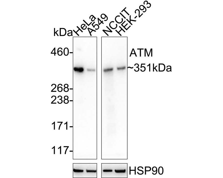
Western blot analysis of ATM on different lysates with Rabbit anti-ATM antibody at 1/5,000 dilution. Lane 1: HeLa cell lysate, Lane 2: A549 cell lysate, Lane 3: NCCIT cell lysate, Lane 4: HEK-293 cell lysate, Lysates/proteins at 15 µg/Lane. Exposure time: 24 seconds; 3-8% SDS-PAGE gel. Proteins were transferred to a PVDF membrane and blocked with 5% NFDM/TBST for 1 hour at room temperature. The primary antibody at 1/5,000 dilution was used in 5% NFDM/TBST at 4℃ overnight. Goat Anti-Rabbit IgG - HRP Secondary Antibody at 1/50,000 dilution was used for 1 hour at room temperature.WB
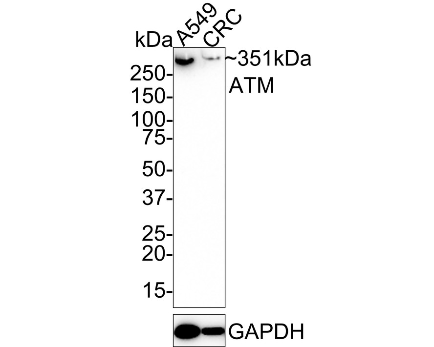
Western blot analysis of ATM on different lysates with Rabbit anti-ATM antibody at 1/1,000 dilution. Lane 1: A549 cell lysate, Lane 2: CRC cell lysate, Lysates/proteins at 20 µg/Lane. Exposure time: 3 minutes; 4-20% SDS-PAGE gel. Proteins were transferred to a PVDF membrane and blocked with 5% NFDM/TBST for 1 hour at room temperature. The primary antibody at 1/1,000 dilution was used in 5% NFDM/TBST at 4℃ overnight. Goat Anti-Rabbit IgG - HRP Secondary Antibody at 1/50,000 dilution was used for 1 hour at room temperature.IHC
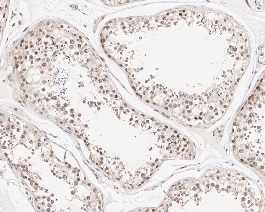
Immunohistochemical analysis of paraffin-embedded human testis tissue with Rabbit anti-ATM antibody at 1/1,000 dilution. The section was pre-treated using heat mediated antigen retrieval with sodium citrate buffer (pH 6.0) for 2 minutes. The tissues were blocked in 1% BSA for 20 minutes at room temperature, washed with ddH2O and PBS, and then probed with the primary antibody at 1/1,000 dilution for 1 hour at room temperature. The detection was performed using an HRP conjugated compact polymer system. DAB was used as the chromogen. Tissues were counterstained with hematoxylin and mounted with DPX.IHC
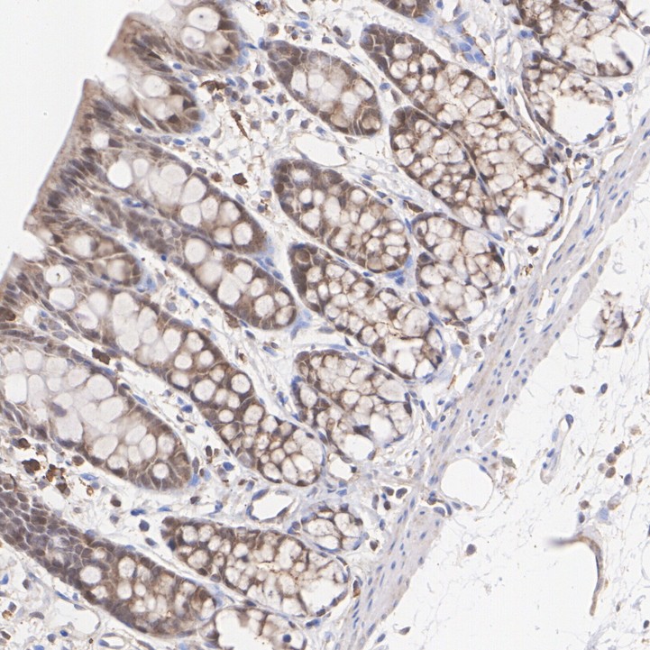
Immunohistochemical analysis of paraffin-embedded rat colon tissue with Rabbit anti-ATM antibody at 1/200 dilution. The section was pre-treated using heat mediated antigen retrieval with sodium citrate buffer (pH 6.0) for 2 minutes. The tissues were blocked in 1% BSA for 20 minutes at room temperature, washed with ddH2O and PBS, and then probed with the primary antibody at 1/200 dilution for 1 hour at room temperature. The detection was performed using an HRP conjugated compact polymer system. DAB was used as the chromogen. Tissues were counterstained with hematoxylin and mounted with DPX.ICC/IF
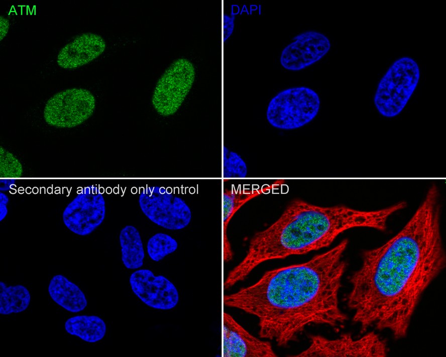
Immunocytochemistry analysis of HeLa cells labeling ATM with Rabbit anti-ATM antibody at 1/100 dilution. Cells were fixed in 4% paraformaldehyde for 15 minutes at room temperature, permeabilized with 0.1% Triton X-100 in PBS for 15 minutes at room temperature, then blocked with 1% BSA in 10% negative goat serum for 1 hour at room temperature. Cells were then incubated with Rabbit anti-ATM antibody at 1/100 dilution in 1% BSA in PBST overnight at 4 ℃. Goat Anti-Rabbit IgG H&L (488) was used as the secondary antibody at 1/1,000 dilution. PBS instead of the primary antibody was used as the secondary antibody only control. Nuclear DNA was labelled in blue with DAPI. Beta tubulin (red) was stained at 1/100 dilution overnight at +4℃. Goat Anti-Mouse IgG H&L (594) was used as the secondary antibody at 1/1,000 dilution.| Product Name | ATM Recombinant Rabbit Monoclonal Antibody |
|---|---|
| Antibody Type | Primary Antibodies |
| Product description | The phosphatidylinositol kinase (PIK) family members fall into two distinct subgroups. The first subgroup contains proteins such as the PI 3- and PI 4-kinases and the second group comprises the PIK-related kinases. The PIK-related kinases include Atm, DNA-PKCS and FRAP. These proteins have in common a region of homology at their carboxy-termini that is not present in the PI 3- and PI 4-kinases. The Atm gene is mutated in the autosomal recessive disorder ataxia telangiectasia (AT) that is characterized by cerebellar degeneration (ataxia) and the appearance of dilated blood vessels (telangiec-tases) in the conjunctivae of the eyes. AT cells are hypersensitive to ionizing radiation, impaired in mediating the inhibition of DNA synthesis and display delays in p53 induction. |
| Immunogen | Synthetic peptide within Human ATM aa 1,951-2,000 / 3,056. |
| Clonality | monoclonal |
|---|---|
| Isotype | IgG |
| Host Species | Recombinant rabbit |
| Tested Applications | ICC/IFIHCWB |
| WB:1:1000-1:5000 IHC:1:50-1:1000 ICC/IF:1:50-1:200 |
|
| Species Reactivity | HumanRat |
| Concentration | 1mg/ml |
| Purification | Protein A |
| Gene Symbol | ATM |
|---|---|
| Gene Synonyms | AT1 ATA ATC ATD ATE ATDC TEL1 TELO1 |
| Gene Full Name | ATM serine/threonine kinase |
| Gene Summary | The protein encoded by this gene belongs to the PI3/PI4-kinase family. This protein is an important cell cycle checkpoint kinase that phosphorylates; thus, it functions as a regulator of a wide variety of downstream proteins, including tumor suppressor proteins p53 and BRCA1, checkpoint kinase CHK2, checkpoint proteins RAD17 and RAD9, and DNA repair protein NBS1. This protein and the closely related kinase ATR are thought to be master controllers of cell cycle checkpoint signaling pathways that are required for cell response to DNA damage and for genome stability. Mutations in this gene are associated with ataxia telangiectasia, an autosomal recessive disorder. [provided by RefSeq, Aug 2010] |
| Alternative Names | A-T mutated antibody
A-T mutated homolog antibody
AT mutated antibody
AT1 antibody
ATA antibody
Ataxia telangiectasia mutated antibody
Ataxia telangiectasia mutated gene antibody
Ataxia telangiectasia mutated homolog (human) antibody
Ataxia telangiectasia mutated homolog antibody
ATC antibody
ATD antibody
ATDC antibody
ATE antibody
ATM antibody
ATM serine/threonine kinase antibody
ATM_HUMAN antibody
DKFZp781A0353 antibody
MGC74674 antibody
OTTHUMP00000232981 antibody
Serine protein kinase ATM antibody
Serine-protein kinase ATM antibody
Serine/threonine-protein kinase ATM antibody
Tefu antibody
TEL1 antibody
TEL1 telomere maintenance 1 homolog antibody TELO1 antibody Telomere fusion protein antibody |
| Molecular Weight(MW) | 351kDa |
| Cellular Localization | Nucleus, Cytoplasmic vesicle. |
| SwissProt ID | Q13315 |
|---|
WB
Western blot analysis of ATM on different lysates with Rabbit anti-ATM antibody at 1/5,000 dilution. Lane 1: HeLa cell lysate, Lane 2: A549 cell lysate, Lane 3: NCCIT cell lysate, Lane 4: HEK-293 cell lysate, Lysates/proteins at 15 µg/Lane. Exposure time: 24 seconds; 3-8% SDS-PAGE gel. Proteins were transferred to a PVDF membrane and blocked with 5% NFDM/TBST for 1 hour at room temperature. The primary antibody at 1/5,000 dilution was used in 5% NFDM/TBST at 4℃ overnight. Goat Anti-Rabbit IgG - HRP Secondary Antibody at 1/50,000 dilution was used for 1 hour at room temperature.
WB
Western blot analysis of ATM on different lysates with Rabbit anti-ATM antibody at 1/1,000 dilution. Lane 1: A549 cell lysate, Lane 2: CRC cell lysate, Lysates/proteins at 20 µg/Lane. Exposure time: 3 minutes; 4-20% SDS-PAGE gel. Proteins were transferred to a PVDF membrane and blocked with 5% NFDM/TBST for 1 hour at room temperature. The primary antibody at 1/1,000 dilution was used in 5% NFDM/TBST at 4℃ overnight. Goat Anti-Rabbit IgG - HRP Secondary Antibody at 1/50,000 dilution was used for 1 hour at room temperature.
IHC
Immunohistochemical analysis of paraffin-embedded human testis tissue with Rabbit anti-ATM antibody at 1/1,000 dilution. The section was pre-treated using heat mediated antigen retrieval with sodium citrate buffer (pH 6.0) for 2 minutes. The tissues were blocked in 1% BSA for 20 minutes at room temperature, washed with ddH2O and PBS, and then probed with the primary antibody at 1/1,000 dilution for 1 hour at room temperature. The detection was performed using an HRP conjugated compact polymer system. DAB was used as the chromogen. Tissues were counterstained with hematoxylin and mounted with DPX.
IHC
Immunohistochemical analysis of paraffin-embedded rat colon tissue with Rabbit anti-ATM antibody at 1/200 dilution. The section was pre-treated using heat mediated antigen retrieval with sodium citrate buffer (pH 6.0) for 2 minutes. The tissues were blocked in 1% BSA for 20 minutes at room temperature, washed with ddH2O and PBS, and then probed with the primary antibody at 1/200 dilution for 1 hour at room temperature. The detection was performed using an HRP conjugated compact polymer system. DAB was used as the chromogen. Tissues were counterstained with hematoxylin and mounted with DPX.
ICC/IF
Immunocytochemistry analysis of HeLa cells labeling ATM with Rabbit anti-ATM antibody at 1/100 dilution. Cells were fixed in 4% paraformaldehyde for 15 minutes at room temperature, permeabilized with 0.1% Triton X-100 in PBS for 15 minutes at room temperature, then blocked with 1% BSA in 10% negative goat serum for 1 hour at room temperature. Cells were then incubated with Rabbit anti-ATM antibody at 1/100 dilution in 1% BSA in PBST overnight at 4 ℃. Goat Anti-Rabbit IgG H&L (488) was used as the secondary antibody at 1/1,000 dilution. PBS instead of the primary antibody was used as the secondary antibody only control. Nuclear DNA was labelled in blue with DAPI. Beta tubulin (red) was stained at 1/100 dilution overnight at +4℃. Goat Anti-Mouse IgG H&L (594) was used as the secondary antibody at 1/1,000 dilution.| Positive Control | CRC, MCF-7, Hela, human liver cancer tissue, human tonsil tissue, human pancreas tissue. |
|---|---|
| Application Notes | WB:1:1000-1:5000 IHC:1:50-1:1000 ICC/IF:1:50-1:200 |
| Form | Liquid |
|---|---|
| Storage Instructions | Store at +4℃ after thawing. Aliquot store at -20℃ or -80℃. Avoid repeated freeze / thaw cycles. |
| Storage Buffer | 1*TBS (pH7.4), 1%BSA, 40%Glycerol. Preservative: 0.05% Sodium Azide. |


