WB
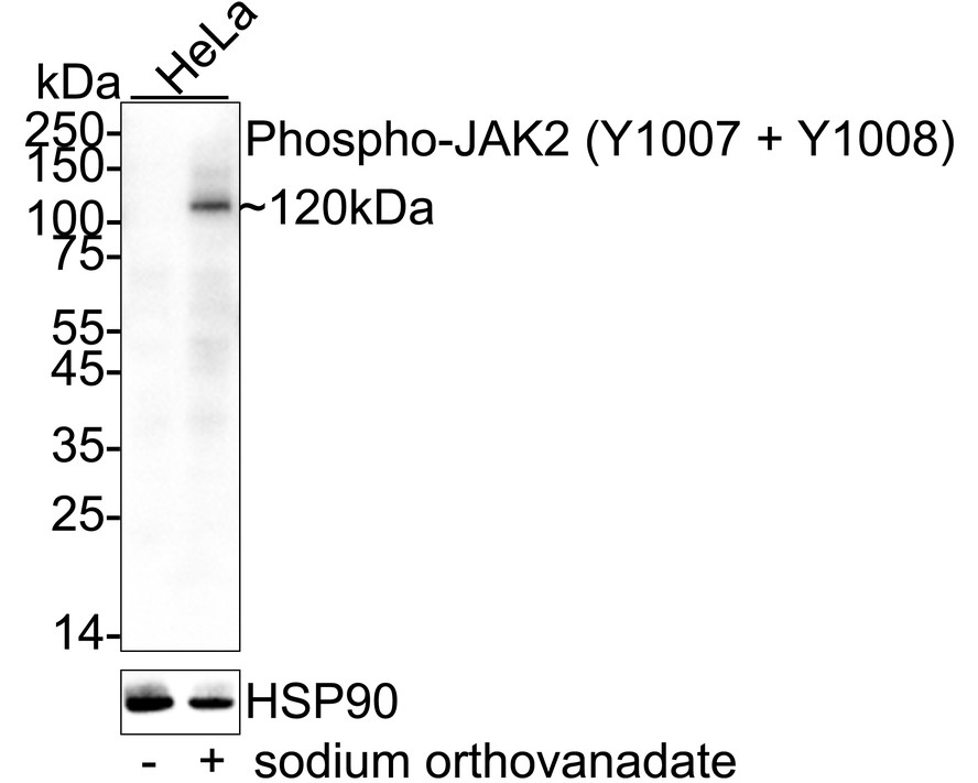
Western blot analysis of Phospho-JAK2 (Y1007 + Y1008) on different lysates with Rabbit anti-Phospho-JAK2 (Y1007 + Y1008) antibody at 1/1,000 dilution. Lane 1: HeLa cell lysate, Lane 2: HeLa treated with 1mM sodium orthovanadate for 30 minutes cell lysate, Lysates/proteins at 20 µg/Lane. Exposure time: 3 minute; 4-20% SDS-PAGE gel. Proteins were transferred to a PVDF membrane and blocked with 5% NFDM/TBST for 1 hour at room temperature. The primary antibody at 1/1,000 dilution was used in 5% NFDM/TBST at 4℃ overnight. Goat Anti-Rabbit IgG - HRP Secondary Antibody at 1/50,000 dilution was used for 1 hour at room temperature.ICC/IF
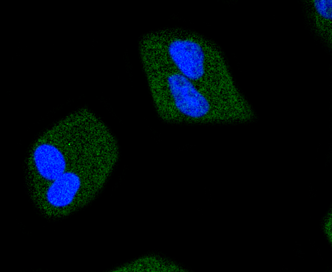
ICC staining of Phospho-JAK2 (Y1007 + Y1008) in Hela cells (green). Formalin fixed cells were permeabilized with 0.1% Triton X-100 in TBS for 10 minutes at room temperature and blocked with 1% Blocker BSA for 15 minutes at room temperature. Cells were probed with the primary antibody (1/50) for 1 hour at room temperature, washed with PBS. Alexa Fluor®488 Goat anti-Rabbit IgG was used as the secondary antibody at 1/1,000 dilution. The nuclear counter stain is DAPI (blue).IHC
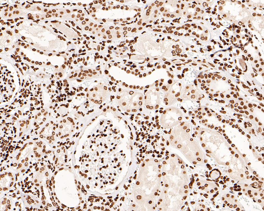
Immunohistochemical analysis of paraffin-embedded human kidney tissue with Rabbit anti-Phospho-JAK2 (Y1007 + Y1008) antibody at 1/500 dilution. The section was pre-treated using heat mediated antigen retrieval with sodium citrate buffer (pH 6.0) for 2 minutes. The tissues were blocked in 1% BSA for 20 minutes at room temperature, washed with ddH2O and PBS, and then probed with the primary antibody at 1/500 dilution for 1 hour at room temperature. The detection was performed using an HRP conjugated compact polymer system. DAB was used as the chromogen. Tissues were counterstained with hematoxylin and mounted with DPX.IHC
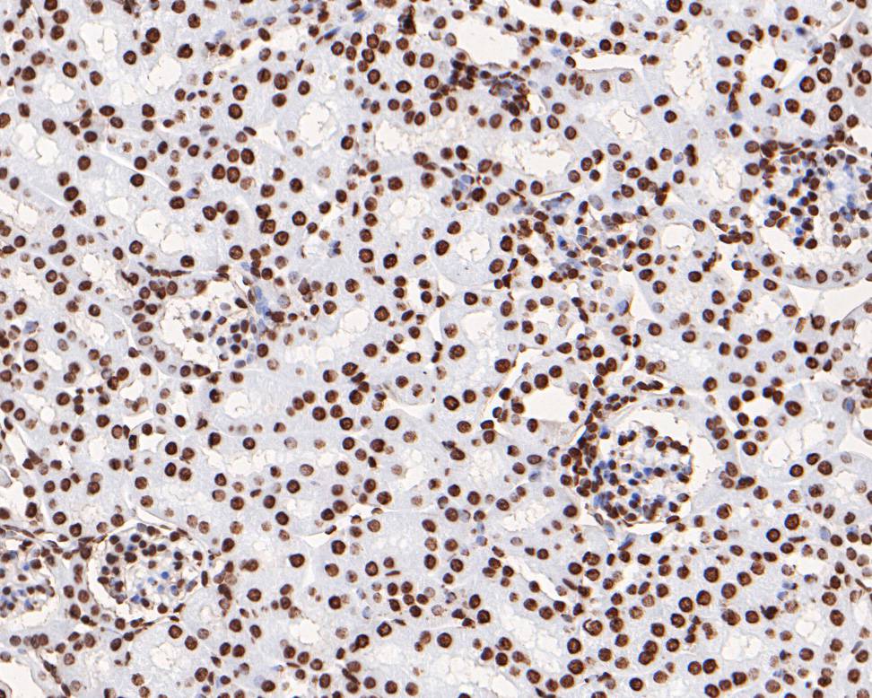
Immunohistochemical analysis of paraffin-embedded mouse kidney tissue using anti-Phospho-JAK2 (Y1007 + Y1008) antibody. The section was pre-treated using heat mediated antigen retrieval with sodium citrate buffer (pH 6.0) for 20 minutes. The tissues were blocked in 5% BSA for 30 minutes at room temperature, washed with ddH2O and PBS, and then probed with the primary antibody (1/200) for 30 minutes at room temperature. The detection was performed using an HRP conjugated compact polymer system. DAB was used as the chromogen. Tissues were counterstained with hematoxylin and mounted with DPX.WB
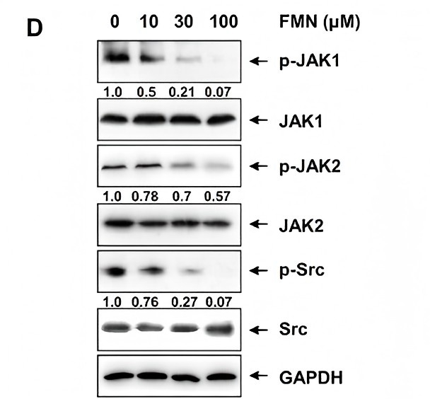
(D) HeLa cells were treated with FMN for 12 h. Whole cell extracts of STAT3 upstream kinase were assessed by western blot.| Product Name | Phospho-JAK2 (Y1007 + Y1008) Recombinant Rabbit Monoclonal Antibody |
|---|---|
| Antibody Type | Primary Antibodies |
| Product description | JAK2 (Janus Kinase 2) belongs to the emerging family of non-receptor Janus tyrosine kinases, which regulate a spectrum of cellular functions downstream of activated cytokine receptors in the lympho-hematopoietic system. Immuno-logical stimuli, such as interferons and cytokines, induce recruitment of Stat transcription factors to cytokine receptor-associated JAK2. JAK2 then phosphorylates proximal Stat factors, which subsequently dimerize, translocate to the nucleus and bind to cis elements upstream of target gene promoters to regulate transcription. The canonical JAK-Stat pathway is integral to maintaining a normal immune system by stimulating proliferation, differentiation, survival, and host resistance to pathogens. Altering JAK-Stat signaling to reduce cytokine induced pro-inflammatory responses represents an attractive target for anti-inflammatory therapies. Within the JAK2 kinase domain, there is a region that has considerable sequence homology to the regulatory region of the insulin receptor. Among a variety of sites, Tyrosines 1007 and 1008 are sites of trans- or autophosphorylation in vivo and in in vitro kinase reactions. |
| Immunogen | Synthetic phospho-peptide corresponding to residues surrounding Tyr1007 and 1008 of human JAK2. |
| Modification | p-Y1007 + Y1008 |
| Clonality | monoclonal |
|---|---|
| Isotype | IgG |
| Host Species | Recombinant rabbit |
| Tested Applications | ICC/IFIHCWB |
| WB:1:1000 ICC/IF:1:50-1:200 IHC:1:200-1:500 |
|
| Species Reactivity | HumanMouseRat |
| Concentration | 1mg/ml |
| Purification | Protein A |
| Gene Symbol | JAK2 |
|---|---|
| Gene Synonyms | JTK10 |
| Gene Full Name | Janus kinase 2 |
| Molecular Weight(MW) | 131kDa(Observed band size: 120kDa) |
| Cellular Localization | Nucleus, Endomembrane system, Cytoplasm. |
WB
Western blot analysis of Phospho-JAK2 (Y1007 + Y1008) on different lysates with Rabbit anti-Phospho-JAK2 (Y1007 + Y1008) antibody at 1/1,000 dilution. Lane 1: HeLa cell lysate, Lane 2: HeLa treated with 1mM sodium orthovanadate for 30 minutes cell lysate, Lysates/proteins at 20 µg/Lane. Exposure time: 3 minute; 4-20% SDS-PAGE gel. Proteins were transferred to a PVDF membrane and blocked with 5% NFDM/TBST for 1 hour at room temperature. The primary antibody at 1/1,000 dilution was used in 5% NFDM/TBST at 4℃ overnight. Goat Anti-Rabbit IgG - HRP Secondary Antibody at 1/50,000 dilution was used for 1 hour at room temperature.
ICC/IF
ICC staining of Phospho-JAK2 (Y1007 + Y1008) in Hela cells (green). Formalin fixed cells were permeabilized with 0.1% Triton X-100 in TBS for 10 minutes at room temperature and blocked with 1% Blocker BSA for 15 minutes at room temperature. Cells were probed with the primary antibody (1/50) for 1 hour at room temperature, washed with PBS. Alexa Fluor®488 Goat anti-Rabbit IgG was used as the secondary antibody at 1/1,000 dilution. The nuclear counter stain is DAPI (blue).
IHC
Immunohistochemical analysis of paraffin-embedded human kidney tissue with Rabbit anti-Phospho-JAK2 (Y1007 + Y1008) antibody at 1/500 dilution. The section was pre-treated using heat mediated antigen retrieval with sodium citrate buffer (pH 6.0) for 2 minutes. The tissues were blocked in 1% BSA for 20 minutes at room temperature, washed with ddH2O and PBS, and then probed with the primary antibody at 1/500 dilution for 1 hour at room temperature. The detection was performed using an HRP conjugated compact polymer system. DAB was used as the chromogen. Tissues were counterstained with hematoxylin and mounted with DPX.
IHC
Immunohistochemical analysis of paraffin-embedded mouse kidney tissue using anti-Phospho-JAK2 (Y1007 + Y1008) antibody. The section was pre-treated using heat mediated antigen retrieval with sodium citrate buffer (pH 6.0) for 20 minutes. The tissues were blocked in 5% BSA for 30 minutes at room temperature, washed with ddH2O and PBS, and then probed with the primary antibody (1/200) for 30 minutes at room temperature. The detection was performed using an HRP conjugated compact polymer system. DAB was used as the chromogen. Tissues were counterstained with hematoxylin and mounted with DPX.
WB
(D) HeLa cells were treated with FMN for 12 h. Whole cell extracts of STAT3 upstream kinase were assessed by western blot.| Positive Control | Hela, human tonsil tissue, human spleen tissue, mouse spleen tissue. |
|---|---|
| Application Notes | WB:1:1000 ICC/IF:1:50-1:200 IHC:1:200-1:500 |
| Form | Liquid |
|---|---|
| Storage Instructions | Store at +4℃ after thawing. Aliquot store at -20℃ or -80℃. Avoid repeated freeze / thaw cycles. |
| Storage Buffer | 1*TBS (pH7.4), 1%BSA, 40%Glycerol. Preservative: 0.05% Sodium Azide. |


