Application
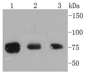
Fig1: Western blot analysis of Phospho-RSK1 p90(T359+S363) on different lysates using anti-Phospho-RSK1 p90(T359+S363) antibody at 1/1,000 dilution. Positive control: Lane 1: A431 Lane 2: 293 Lane 3: MCF-7Application
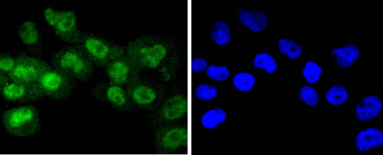
Fig2: ICC staining Phospho-RSK1 p90(T359+S363) in A431 cells (green). The nuclear counter stain is DAPI (blue). Cells were fixed in paraformaldehyde, permeabilised with 0.25% Triton X100/PBS.Application
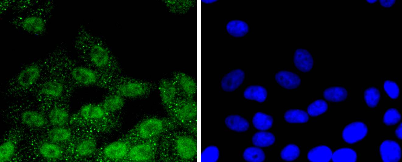
Fig3: ICC staining Phospho-RSK1 p90(T359+S363) in HepG2 cells (green). The nuclear counter stain is DAPI (blue). Cells were fixed in paraformaldehyde, permeabilised with 0.25% Triton X100/PBS.Application
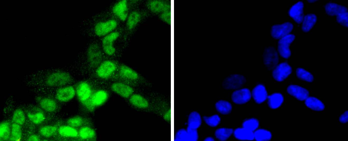
Fig4: ICC staining Phospho-RSK1 p90(T359+S363) in 293 cells (green). The nuclear counter stain is DAPI (blue). Cells were fixed in paraformaldehyde, permeabilised with 0.25% Triton X100/PBS.Application
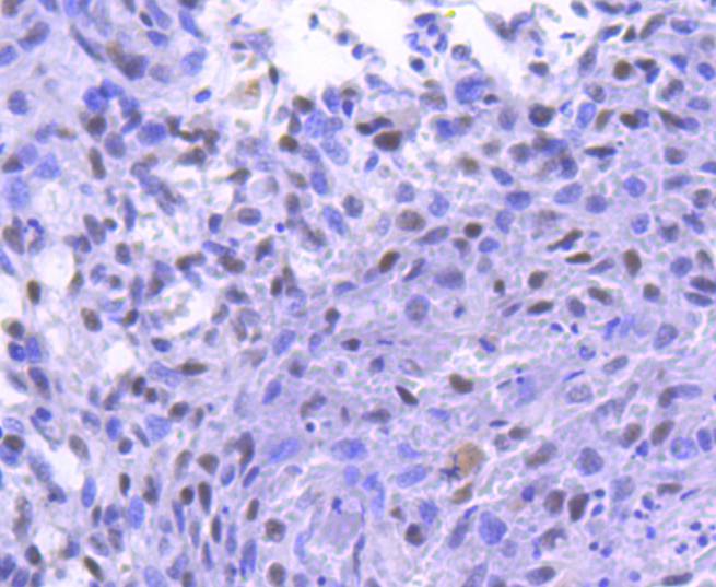
Fig5: Immunohistochemical analysis of paraffin-embedded human breast carcinoma tissue using anti-Phospho-RSK1 p90(T359+S363) antibody. Counter stained with hematoxylin.Application
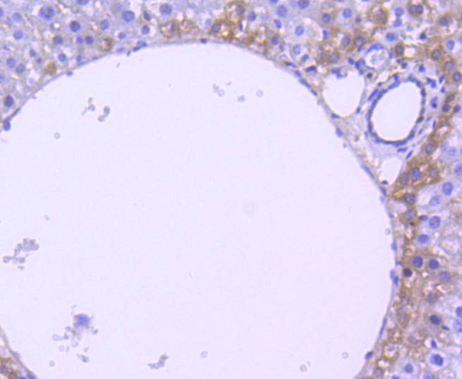
Fig6: Immunohistochemical analysis of paraffin-embedded mouse liver tissue using anti-Phospho-RSK1 p90(T359+S363) antibody. Counter stained with hematoxylin.| Product Name | Phospho-RSK1 p90(T359+S363) [SU03-65] |
|---|---|
| Antibody Type | Primary Antibodies |
| Antigen Alias | 90 kDa ribosomal protein S6 kinase 1 antibody dJ590P13.1 (ribosomal protein S6 kinase, 90kD, polypeptide 1 antibody dJ590P13.1 antibody EC 2.7.11.1 antibody HU 1 antibody HU1 antibody KS6A1_HUMAN antibody MAP kinase activated protein kinase 1a antibody MAP kinase-activated protein kinase 1a antibody MAPK-activated protein kinase 1a antibody MAPKAP kinase 1a antibody MAPKAPK-1a antibody MAPKAPK1A antibody MGC79981 antibody Mitogen-activated protein kinase-activated protein kinase 1A antibody OTTHUMP00000004113 antibody p90 RSK1 antibody p90-RSK 1 antibody p90rsk antibody p90RSK1 antibody p90S6K antibody pp90RSK1 antibody Ribosomal protein S6 kinase 90kD 1 antibody Ribosomal protein S6 kinase 90kD polypeptide 1 antibody Ribosomal protein S6 kinase 90kDa polypeptide 1 antibody Ribosomal protein S6 kinase alpha 1 antibody Ribosomal protein S6 kinase alpha-1 antibody Ribosomal protein S6 kinase polypeptide 1 antibody Ribosomal S6 kinase 1 antibody RPS6K1 alpha antibody rps6ka antibody Rps6ka1 antibody RSK 1 antibody RSK 1 p90 antibody RSK antibody RSK-1 antibody RSK1 antibody S6K alpha 1 antibody S6K-alpha-1 antibody |
| Product description | The family of ribosomal S6 kinases (Rsks), designated Rsk-1 (or MAPKAP kinase-1), Rsk-2 and Rsk-3, are intracellular serine/threonine kinases that are important signaling intermediates in response to a broad range of ligand activated receptor tyrosine kinases. A unique feature common to the members of the Rsk family is that each possesses two non-identical complete kinase catalytic domains. An additional Rsk protein, Rsk-4, shows a high level of homology to the three previously isolated members of the human Rsk family. Rsk-4 is most abundantly expressed in brain and kidney and plays a role in normal neuronal development. The family of ribosomal S6 kinases includes p70 S6 kinase and p70 S6 kinase β, which are thought to have similar regulatory functions. MSK1 (also designated RLPK) is a novel Rsk-related protein, which, like the p90 Rsk family members, contains two non-identical complete kinase catalytic domains. |
| Immunogen | Synthetic phospho-peptide corresponding to residues surrounding Thr359 and Ser363 of human RSK1 p90 |
| Modification | p-T359+S363 |
| Clonality | Monoclonal |
|---|---|
| Isotype | IgG |
| Host Species | Recombinant rabbit |
| Tested Applications | WBICCIHCIP |
| WB:1:1,000-1:2,000 ICC:1:50-1:200 IHC:1:50-1:200 |
|
| Species Reactivity | HumanMouseRat |
| Concentration | 1mg/ml |
| Alternative Names | 90 kDa ribosomal protein S6 kinase 1 antibody
dJ590P13.1 (ribosomal protein S6 kinase 90kD polypeptide 1 antibody dJ590P13.1 antibody EC 2.7.11.1 antibody HU 1 antibody HU1 antibody KS6A1_HUMAN antibody MAP kinase activated protein kinase 1a antibody MAP kinase-activated protein kinase 1a antibody MAPK-activated protein kinase 1a antibody MAPKAP kinase 1a antibody MAPKAPK-1a antibody MAPKAPK1A antibody MGC79981 antibody Mitogen-activated protein kinase-activated protein kinase 1A antibody OTTHUMP00000004113 antibody p90 RSK1 antibody p90-RSK 1 antibody p90rsk antibody p90RSK1 antibody p90S6K antibody pp90RSK1 antibody Ribosomal protein S6 kinase 90kD 1 antibody Ribosomal protein S6 kinase 90kD polypeptide 1 antibody Ribosomal protein S6 kinase 90kDa polypeptide 1 antibody Ribosomal protein S6 kinase alpha 1 antibody Ribosomal protein S6 kinase alpha-1 antibody Ribosomal protein S6 kinase polypeptide 1 antibody Ribosomal S6 kinase 1 antibody RPS6K1 alpha antibody rps6ka antibody Rps6ka1 antibody RSK 1 antibody RSK 1 p90 antibody RSK antibody RSK-1 antibody RSK1 antibody S6K alpha 1 antibody S6K-alpha-1 antibody |
|---|---|
| Molecular Weight(MW) | 90 kDa |
| Cellular Localization | Nucleus, Cytoplasm. |
Application
Fig1: Western blot analysis of Phospho-RSK1 p90(T359+S363) on different lysates using anti-Phospho-RSK1 p90(T359+S363) antibody at 1/1,000 dilution. Positive control: Lane 1: A431 Lane 2: 293 Lane 3: MCF-7
Application
Fig2: ICC staining Phospho-RSK1 p90(T359+S363) in A431 cells (green). The nuclear counter stain is DAPI (blue). Cells were fixed in paraformaldehyde, permeabilised with 0.25% Triton X100/PBS.
Application
Fig3: ICC staining Phospho-RSK1 p90(T359+S363) in HepG2 cells (green). The nuclear counter stain is DAPI (blue). Cells were fixed in paraformaldehyde, permeabilised with 0.25% Triton X100/PBS.
Application
Fig4: ICC staining Phospho-RSK1 p90(T359+S363) in 293 cells (green). The nuclear counter stain is DAPI (blue). Cells were fixed in paraformaldehyde, permeabilised with 0.25% Triton X100/PBS.
Application
Fig5: Immunohistochemical analysis of paraffin-embedded human breast carcinoma tissue using anti-Phospho-RSK1 p90(T359+S363) antibody. Counter stained with hematoxylin.
Application
Fig6: Immunohistochemical analysis of paraffin-embedded mouse liver tissue using anti-Phospho-RSK1 p90(T359+S363) antibody. Counter stained with hematoxylin.| Positive Control | A431, 293, HepG2, mouse liver tissue, human breast carcinoma tissue. |
|---|---|
| Application Notes | WB:1:1,000-1:2,000 ICC:1:50-1:200 IHC:1:50-1:200 |
| Form | Liquid |
|---|---|
| Storage Instructions | Store at +4℃ after thawing. Aliquot store at -20℃ or -80℃. Avoid repeated freeze / thaw cycles. |
| Storage Buffer | 1*TBS (pH7.4), 1%BSA, 40%Glycerol. Preservative: 0.05% Sodium Azide. |


