WB
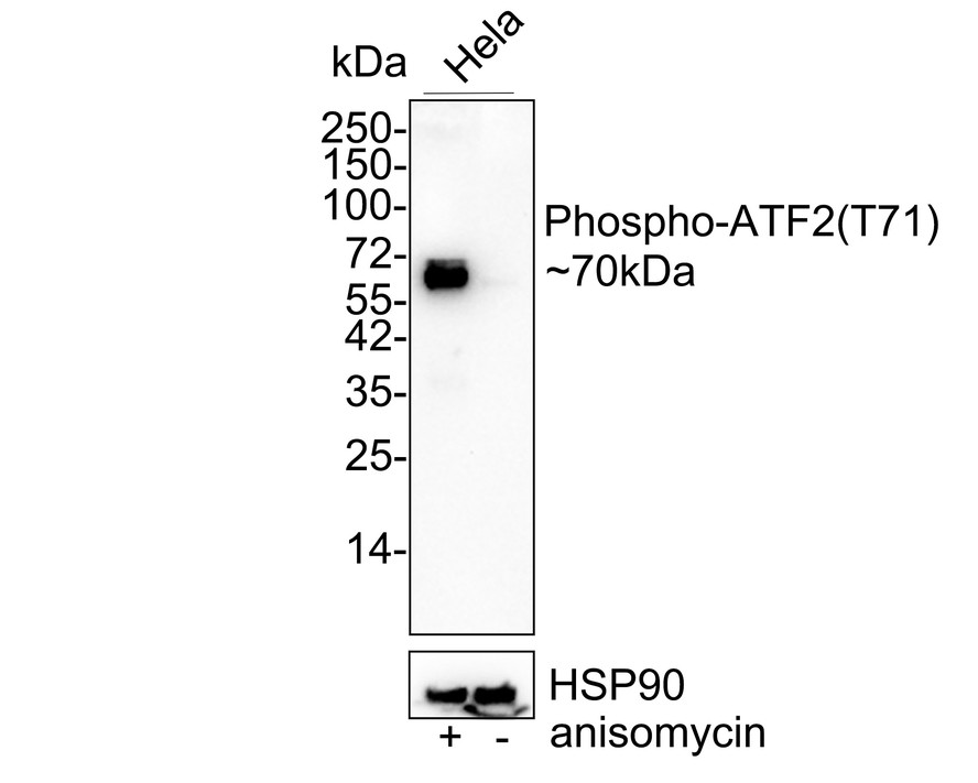
Western blot analysis of Phospho-ATF2 (T71) on HeLa cell lysates treated with or without untreated anisomycin(250ng/ml 30min) with Rabbit anti-Phospho-ATF2 (T71) antibody at 1/1,000 dilution. Lysates/proteins at 20 µg/Lane. Exposure time: 3 minutes; 4-20% SDS-PAGE gel. Proteins were transferred to a PVDF membrane and blocked with 5% NFDM/TBST for 1 hour at room temperature. The primary antibody at 1/1,000 dilution was used in 5% NFDM/TBST at 4℃ overnight. Goat Anti-Rabbit IgG - HRP Secondary Antibody at 1/50,000 dilution was used for 1 hour at room temperature.ICC/IF
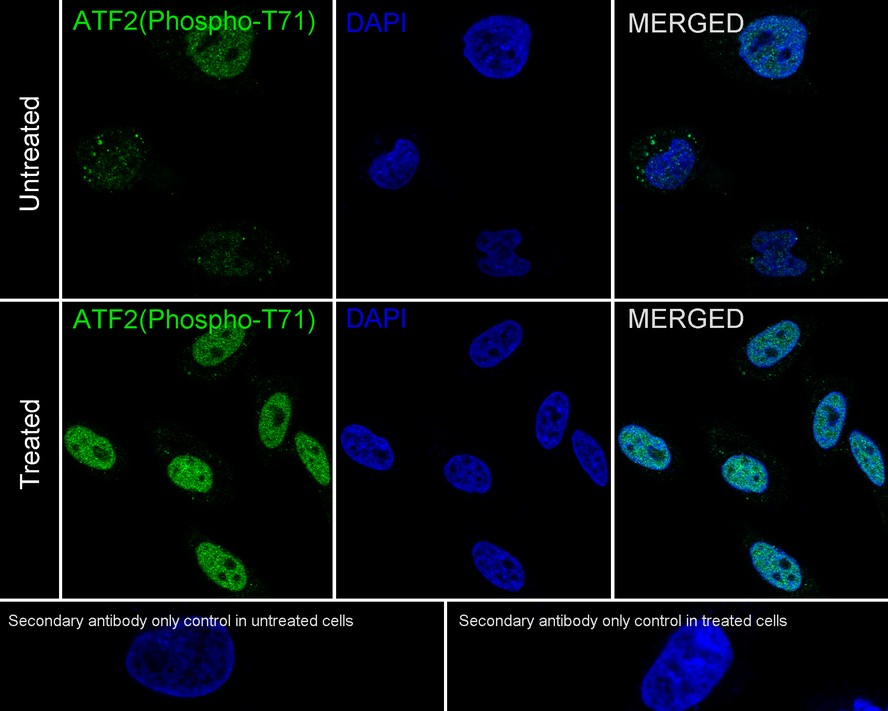
Immunocytochemistry analysis of Hela cells treated with or without 250ng/mL anisomycin for 30 minutes labeling Phospho-ATF2 (T71) with Rabbit anti-Phospho-ATF2 (T71) antibody at 1/50 dilution. Cells were fixed in 4% paraformaldehyde for 10 minutes at 37 ℃, permeabilized with 0.05% Triton X-100 in PBS for 20 minutes, and then blocked with 2% negative goat serum for 30 minutes at room temperature. Cells were then incubated with Rabbit anti-Phospho-ATF2 (T71) antibody at 1/50 dilution in 2% negative goat serum overnight at 4 ℃. Goat Anti-Rabbit IgG H&L (488) was used as the secondary antibody at 1/1,000 dilution. PBS instead of the primary antibody was used as the secondary antibody only control. Nuclear DNA was labelled in blue with DAPI.ICC/IF
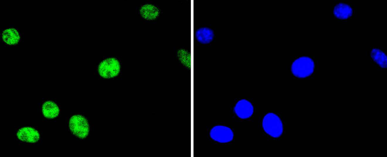
ICC staining phospho-ATF2(T71) in SH-SY-5Y cells (green). The nuclear counter stain is DAPI (blue). Cells were fixed in paraformaldehyde, permeabilised with 0.25% Triton X100/PBS.IHC
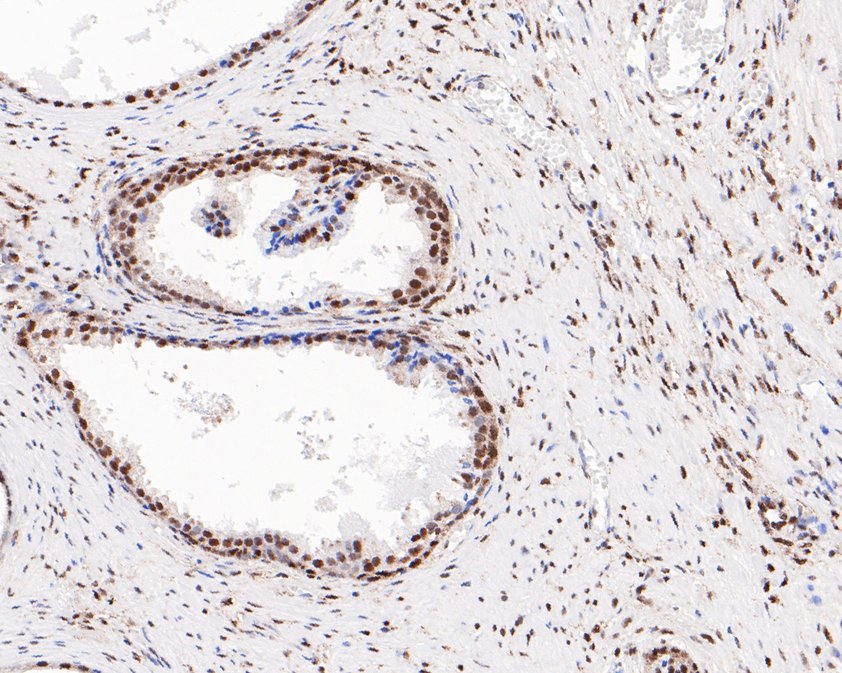
Immunohistochemical analysis of paraffin-embedded human prostate tissue using anti-Phospho-ATF2 (T71) antibody. The section was pre-treated using heat mediated antigen retrieval with sodium citrate buffer (pH 6.0) (high pressure) for 2 minutes. The tissues were blocked in 5% BSA for 30 minutes at room temperature, washed with ddH2O and PBS, and then probed with the primary antibody (1/200) for 30 minutes at room temperature. The detection was performed using an HRP conjugated compact polymer system. DAB was used as the chromogen. Tissues were counterstained with hematoxylin and mounted with DPX.IHC
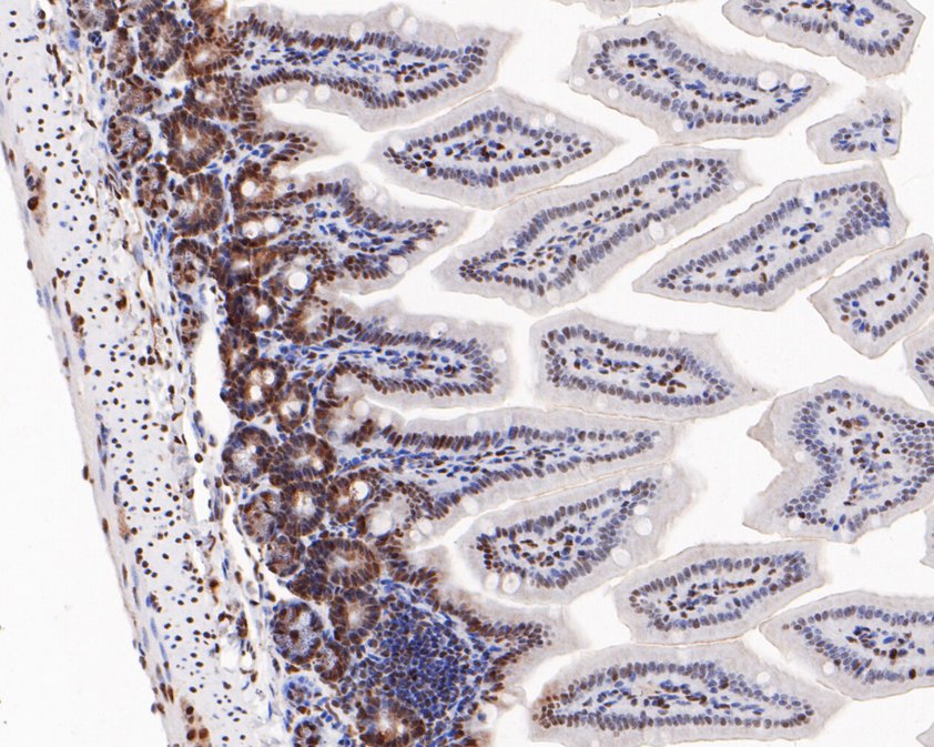
Immunohistochemical analysis of paraffin-embedded mouse colon tissue using anti-Phospho-ATF2 (T71) antibody. The section was pre-treated using heat mediated antigen retrieval with sodium citrate buffer (pH 6.0) (high pressure) for 2 minutes. The tissues were blocked in 5% BSA for 30 minutes at room temperature, washed with ddH2O and PBS, and then probed with the primary antibody (1/200) for 30 minutes at room temperature. The detection was performed using an HRP conjugated compact polymer system. DAB was used as the chromogen. Tissues were counterstained with hematoxylin and mounted with DPX.IHC
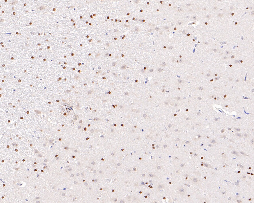
Immunohistochemical analysis of paraffin-embedded rat brain tissue with Rabbit anti-Phospho-ATF2 (T71) antibody at 1/800 dilution. The section was pre-treated using heat mediated antigen retrieval with sodium citrate buffer (pH 6.0) (high pressure) for 2 minutes. The tissues were blocked in 1% BSA for 20 minutes at room temperature, washed with ddH2O and PBS, and then probed with the primary antibody at 1/800 dilution for 1 hour at room temperature. The detection was performed using an HRP conjugated compact polymer system. DAB was used as the chromogen. Tissues were counterstained with hematoxylin and mounted with DPX.IP
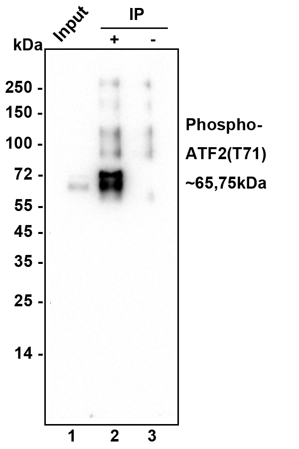
Phospho-ATF2 (T71) was immunoprecipitated in 0.2mg HeLa cell treated anisomycin(25ug/ml 30min) lysate with Rabbit anti-Phospho-ATF2 (T71) antibody at 2 µg/10 µl beads. Western blot was performed from the immunoprecipitate using Rabbit anti-Phospho-ATF2 (T71) antibody at 1/1,000 dilution. Anti-Rabbit IgG for IP Nano-secondary antibody at 1/5,000 dilution was used for 1 hour at room temperature. Lane 1: HeLa cell lysate (input), Lane 2: Rabbit anti-Phospho-ATF2 (T71) antibody IP in HeLa cell lysate, Lane 3: Rabbit IgG instead of Rabbit anti-Phospho-ATF2 (T71) antibody in HeLa cell lysate. Blocking/Dilution buffer: 5% NFDM/TBST. Exposure time: 3 mintues.| Product Name | Phospho-ATF2 (T71) Recombinant Rabbit Monoclonal Antibody |
|---|---|
| Antibody Type | Primary Antibodies |
| Immunogen | Synthetic phospho-peptide corresponding to residues surrounding Thr71 of Human ATF2 aa 58-94 / 505. |
| Modification | p-T71 |
| Clonality | Monoclonal |
|---|---|
| Isotype | IgG |
| Host Species | Recombinant rabbit |
| Tested Applications | ICC/IFIHCIPWB |
| WB:1:1000 IHC:1:100-1:800 ICC/IF:1:50-1:200 |
|
| Species Reactivity | HumanMouseRabbit |
| Concentration | 1mg/ml |
| Purification | Protein A |
| Gene Symbol | ATF2 |
|---|---|
| Gene Synonyms | HB16 CREB2 TREB7 CREB-2 CRE-BP1 |
| Gene Full Name | activating transcription factor 2 |
| Gene Summary | This gene encodes a transcription factor that is a member of the leucine zipper family of DNA binding proteins. The encoded protein has been identified as a moonlighting protein based on its ability to perform mechanistically distinct functions This protein binds to the cAMP-responsive element (CRE), an octameric palindrome. It forms a homodimer or a heterodimer with c-Jun and stimulates CRE-dependent transcription. This protein is also a histone acetyltransferase (HAT) that specifically acetylates histones H2B and H4 in vitro; thus it may represent a class of sequence-specific factors that activate transcription by direct effects on chromatin components. The encoded protein may also be involved in cell's DNA damage response independent of its role in transcriptional regulation. Several alternatively spliced transcript variants have been found for this gene [provided by RefSeq, Jan 2014] |
| Molecular Weight(MW) | 70 kDa |
| Cellular Localization | Mitochondrion outer membrane, Nucleus, Cytoplasm. |
WB
Western blot analysis of Phospho-ATF2 (T71) on HeLa cell lysates treated with or without untreated anisomycin(250ng/ml 30min) with Rabbit anti-Phospho-ATF2 (T71) antibody at 1/1,000 dilution. Lysates/proteins at 20 µg/Lane. Exposure time: 3 minutes; 4-20% SDS-PAGE gel. Proteins were transferred to a PVDF membrane and blocked with 5% NFDM/TBST for 1 hour at room temperature. The primary antibody at 1/1,000 dilution was used in 5% NFDM/TBST at 4℃ overnight. Goat Anti-Rabbit IgG - HRP Secondary Antibody at 1/50,000 dilution was used for 1 hour at room temperature.
ICC/IF
Immunocytochemistry analysis of Hela cells treated with or without 250ng/mL anisomycin for 30 minutes labeling Phospho-ATF2 (T71) with Rabbit anti-Phospho-ATF2 (T71) antibody at 1/50 dilution. Cells were fixed in 4% paraformaldehyde for 10 minutes at 37 ℃, permeabilized with 0.05% Triton X-100 in PBS for 20 minutes, and then blocked with 2% negative goat serum for 30 minutes at room temperature. Cells were then incubated with Rabbit anti-Phospho-ATF2 (T71) antibody at 1/50 dilution in 2% negative goat serum overnight at 4 ℃. Goat Anti-Rabbit IgG H&L (488) was used as the secondary antibody at 1/1,000 dilution. PBS instead of the primary antibody was used as the secondary antibody only control. Nuclear DNA was labelled in blue with DAPI.
ICC/IF
ICC staining phospho-ATF2(T71) in SH-SY-5Y cells (green). The nuclear counter stain is DAPI (blue). Cells were fixed in paraformaldehyde, permeabilised with 0.25% Triton X100/PBS.
IHC
Immunohistochemical analysis of paraffin-embedded human prostate tissue using anti-Phospho-ATF2 (T71) antibody. The section was pre-treated using heat mediated antigen retrieval with sodium citrate buffer (pH 6.0) (high pressure) for 2 minutes. The tissues were blocked in 5% BSA for 30 minutes at room temperature, washed with ddH2O and PBS, and then probed with the primary antibody (1/200) for 30 minutes at room temperature. The detection was performed using an HRP conjugated compact polymer system. DAB was used as the chromogen. Tissues were counterstained with hematoxylin and mounted with DPX.
IHC
Immunohistochemical analysis of paraffin-embedded mouse colon tissue using anti-Phospho-ATF2 (T71) antibody. The section was pre-treated using heat mediated antigen retrieval with sodium citrate buffer (pH 6.0) (high pressure) for 2 minutes. The tissues were blocked in 5% BSA for 30 minutes at room temperature, washed with ddH2O and PBS, and then probed with the primary antibody (1/200) for 30 minutes at room temperature. The detection was performed using an HRP conjugated compact polymer system. DAB was used as the chromogen. Tissues were counterstained with hematoxylin and mounted with DPX.
IHC
Immunohistochemical analysis of paraffin-embedded rat brain tissue with Rabbit anti-Phospho-ATF2 (T71) antibody at 1/800 dilution. The section was pre-treated using heat mediated antigen retrieval with sodium citrate buffer (pH 6.0) (high pressure) for 2 minutes. The tissues were blocked in 1% BSA for 20 minutes at room temperature, washed with ddH2O and PBS, and then probed with the primary antibody at 1/800 dilution for 1 hour at room temperature. The detection was performed using an HRP conjugated compact polymer system. DAB was used as the chromogen. Tissues were counterstained with hematoxylin and mounted with DPX.
IP
Phospho-ATF2 (T71) was immunoprecipitated in 0.2mg HeLa cell treated anisomycin(25ug/ml 30min) lysate with Rabbit anti-Phospho-ATF2 (T71) antibody at 2 µg/10 µl beads. Western blot was performed from the immunoprecipitate using Rabbit anti-Phospho-ATF2 (T71) antibody at 1/1,000 dilution. Anti-Rabbit IgG for IP Nano-secondary antibody at 1/5,000 dilution was used for 1 hour at room temperature. Lane 1: HeLa cell lysate (input), Lane 2: Rabbit anti-Phospho-ATF2 (T71) antibody IP in HeLa cell lysate, Lane 3: Rabbit IgG instead of Rabbit anti-Phospho-ATF2 (T71) antibody in HeLa cell lysate. Blocking/Dilution buffer: 5% NFDM/TBST. Exposure time: 3 mintues.| Application Notes | WB:1:1000 IHC:1:100-1:800 ICC/IF:1:50-1:200 |
|---|
| Form | Liquid |
|---|---|
| Storage Instructions | Shipped at 4℃. Store at +4℃ short term (1-2 weeks). Store at -20℃ long term. |
| Storage Buffer | TBS (pH7.4), 0.05% BSA, 40% Glycerol. Preservative: 0.05% Sodium Azide. |


