WB
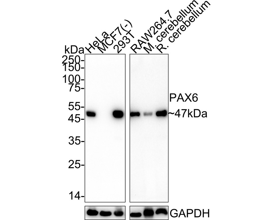
Western blot analysis of PAX6 on different lysates with Rabbit anti-PAX6 antibody at 1/5,000 dilution. Lane 1: HeLa cell lysate, Lane 2: MCF7 cell lysate (negative), Lane 3: 293T cell lysate, Lane 4: RAW264.7 cell lysate, Lane 5: Mouse cerebellum tissue lysate, Lane 6: Rat cerebellum tissue lysate, Lysates/proteins at 20 µg/Lane. Exposure time: Lane 1-3: 1 minute; Lane 4-6: 3 minutes; 4-20% SDS-PAGE gel. Proteins were transferred to a PVDF membrane and blocked with 5% NFDM/TBST for 1 hour at room temperature. The primary antibody at 1/5,000 dilution was used in 5% NFDM/TBST at 4℃ overnight. Goat Anti-Rabbit IgG - HRP Secondary Antibody at 1/50,000 dilution was used for 1 hour at room temperature.IHC
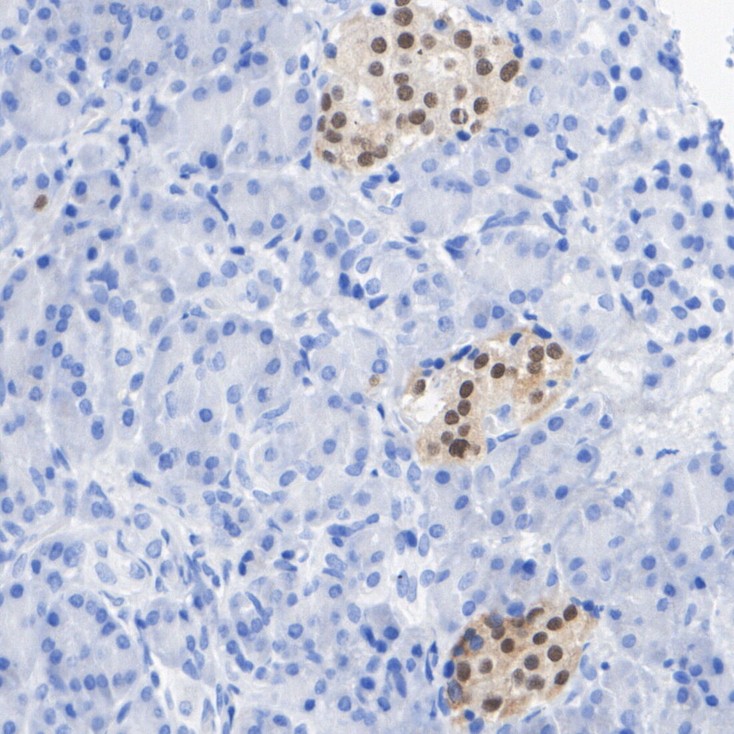
Immunohistochemical analysis of paraffin-embedded human pancreas tissue with Rabbit anti-PAX6 antibody at 1/2,000 dilution. The section was pre-treated using heat mediated antigen retrieval with sodium citrate buffer (pH 6.0) for 2 minutes. The tissues were blocked in 1% BSA for 20 minutes at room temperature, washed with ddH2O and PBS, and then probed with the primary antibody at 1/2,000 dilution for 1 hour at room temperature. The detection was performed using an HRP conjugated compact polymer system. DAB was used as the chromogen. Tissues were counterstained with hematoxylin and mounted with DPX.IHC
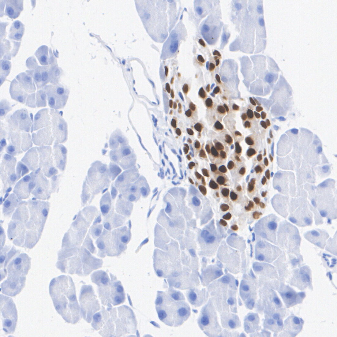
Immunohistochemical analysis of paraffin-embedded mouse pancreas tissue with Rabbit anti-PAX6 antibody at 1/2,000 dilution. The section was pre-treated using heat mediated antigen retrieval with sodium citrate buffer (pH 6.0) for 2 minutes. The tissues were blocked in 1% BSA for 20 minutes at room temperature, washed with ddH2O and PBS, and then probed with the primary antibody at 1/2,000 dilution for 1 hour at room temperature. The detection was performed using an HRP conjugated compact polymer system. DAB was used as the chromogen. Tissues were counterstained with hematoxylin and mounted with DPX.IHC
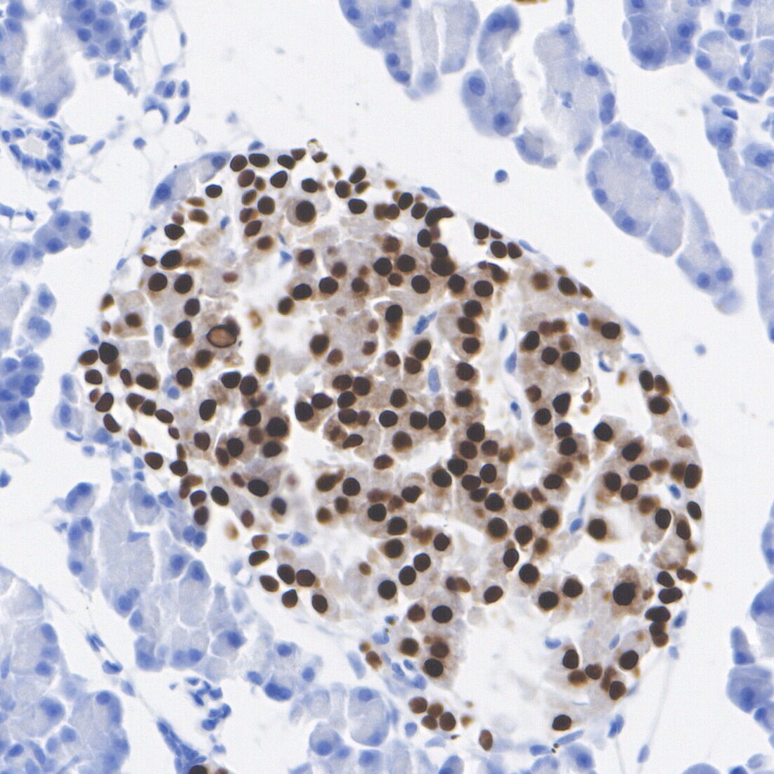
Immunohistochemical analysis of paraffin-embedded rat pancreas tissue with Rabbit anti-PAX6 antibody at 1/2,000 dilution. The section was pre-treated using heat mediated antigen retrieval with sodium citrate buffer (pH 6.0) for 2 minutes. The tissues were blocked in 1% BSA for 20 minutes at room temperature, washed with ddH2O and PBS, and then probed with the primary antibody at 1/2,000 dilution for 1 hour at room temperature. The detection was performed using an HRP conjugated compact polymer system. DAB was used as the chromogen. Tissues were counterstained with hematoxylin and mounted with DPX.ICC/IF
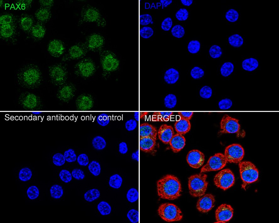
Immunocytochemistry analysis of RAW264.7 cells labeling PAX6 with Rabbit anti-PAX6 antibody at 1/100 dilution. Cells were fixed in 4% paraformaldehyde for 20 minutes at room temperature, permeabilized with 0.1% Triton X-100 in PBS for 5 minutes at room temperature, then blocked with 1% BSA in 10% negative goat serum for 1 hour at room temperature. Cells were then incubated with Rabbit anti-PAX6 antibody at 1/100 dilution in 1% BSA in PBST overnight at 4 ℃. Goat Anti-Rabbit IgG H&L (488) was used as the secondary antibody at 1/1,000 dilution. PBS instead of the primary antibody was used as the secondary antibody only control. Nuclear DNA was labelled in blue with DAPI. Beta tubulin (red) was stained at 1/100 dilution overnight at +4℃. Goat Anti-Mouse IgG H&L (594) was used as the secondary antibody at 1/1,000 dilution.IF-P
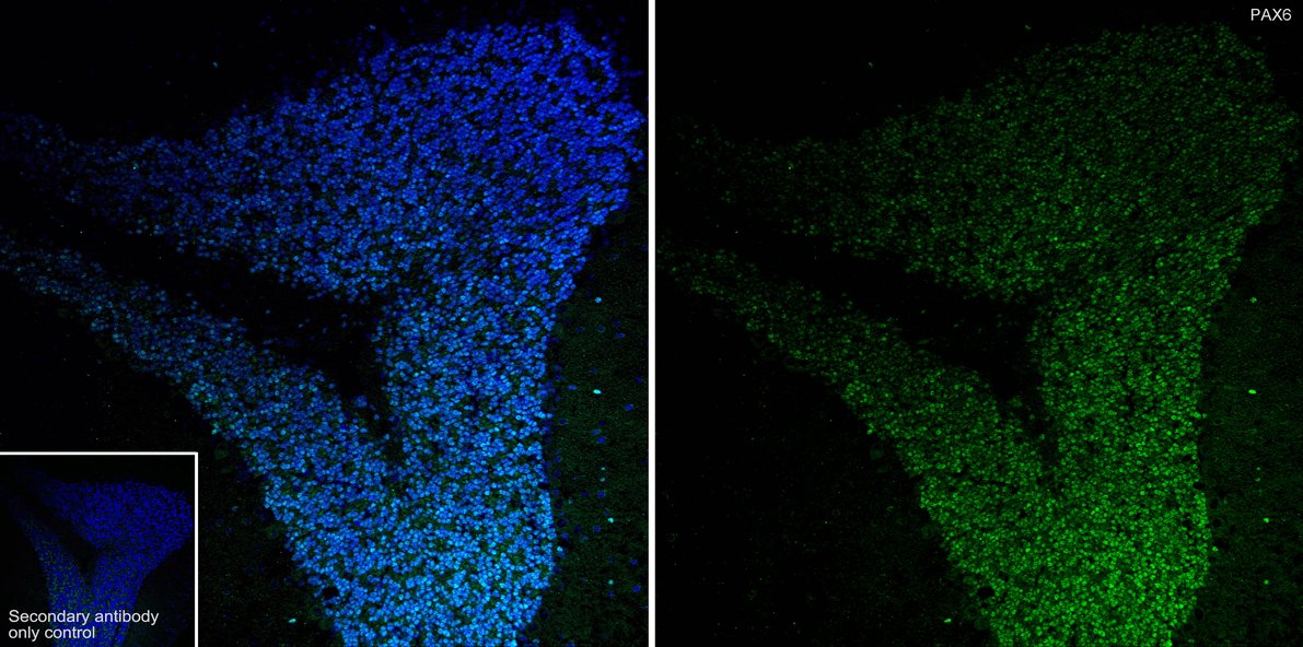
Immunofluorescence analysis of paraffin-embedded mouse cerebellum tissue labeling PAX6 with Rabbit anti-PAX6 antibody at 1/800 dilution. The section was pre-treated using heat mediated antigen retrieval with sodium citrate buffer (pH 6.0) for 2 minutes. The tissues were blocked in 10% negative goat serum for 1 hour at room temperature, washed with PBS, and then probed with the primary antibody (green) at 1/800 dilution overnight at 4 ℃, washed with PBS. Goat Anti-Rabbit IgG H&L (488) was used as the secondary antibody at 1/1,000 dilution. Nuclei were counterstained with DAPI (blue).FC
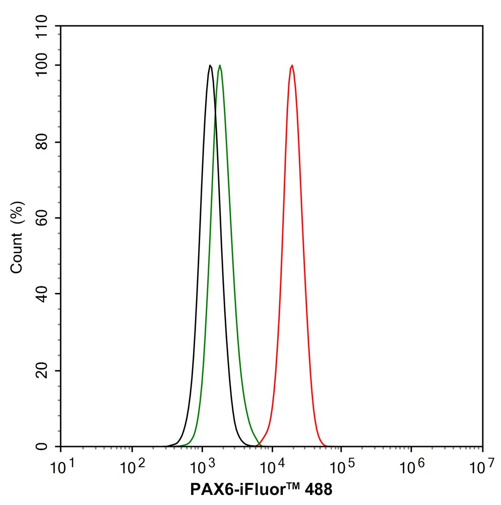
Flow cytometric analysis of RAW264.7 cells labeling PAX6. Cells were fixed and permeabilized. Then stained with the primary antibody (1μg/mL) (red) compared with Rabbit IgG Isotype Control (green). After incubation of the primary antibody at +4℃ for an hour, the cells were stained with a iFluor™ 488 conjugate-Goat anti-Rabbit IgG Secondary antibody at 1/1,000 dilution for 30 minutes at +4℃. Unlabelled sample was used as a control (cells without incubation with primary antibody; black).| Product Name | PAX6 Recombinant Rabbit Monoclonal Antibody |
|---|---|
| Antibody Type | Primary Antibodies |
| Product description | Pax genes contain paired domains with strong homology to genes in Drosophila which are involved in programming early development. Lesions in the Pax-6 gene account for most cases of aniridia, a congenital malformation of the eye, chiefly characterized by iris hypoplasia, which can cause blindness. Pax-6 is involved in other anterior segment malformations besides aniridia, such as Peters anomaly, a major error in the embryonic development of the eye with corneal clouding with variable iridolenticulocorneal adhesions. The Pax-6 gene encodes a transcriptional regulator that recognizes target genes through its paired-type DNA-binding domain. The paired domain is composed of two distinct DNA-binding subdomains, the amino-terminal subdomain and the carboxy-terminal subdomain, which bind respective consensus DNA sequences. The human Pax-6 gene produces two alternatively spliced isoforms that have the distinct structure of the paired domain. |
| Immunogen | Synthetic peptide within Human PAX6 aa 373-422 / 422. |
| Clonality | Monoclonal |
|---|---|
| Isotype | IgG |
| Host Species | Recombinant rabbit |
| Tested Applications | FCICC/IFIF-PIHCWB |
| WB:1:5000 IHC:1:200-1:2000 ICC/IF:1:100 IF-P:1:800 FC:1:1000 |
|
| Species Reactivity | HumanMouseRat |
| Concentration | 1mg/ml |
| Purification | Protein A |
| Gene Symbol | PAX6 |
|---|---|
| Gene Synonyms | AN AN1 AN2 FVH1 MGDA WAGR ASGD5 D11S812E |
| Gene Full Name | paired box 6 |
| Gene Summary | This gene encodes paired box protein Pax-6, one of many human homologs of the Drosophila melanogaster gene prd. In addition to a conserved paired box domain, a hallmark feature of this gene family, the encoded protein also contains a homeobox domain. Both domains are known to bind DNA and function as regulators of gene transcription. Activity of this protein is key in the development of neural tissues, particularly the eye. This gene is regulated by multiple enhancers located up to hundreds of kilobases distant from this locus. Mutations in this gene or in the enhancer regions can cause ocular disorders such as aniridia and Peter's anomaly. Use of alternate promoters and alternative splicing results in multiple transcript variants encoding different isoforms. Interestingly, inclusion of a particular alternate coding exon has been shown to increase the length of the paired box domain and alter its DNA binding specificity. Consequently, isoforms that carry the shorter paired box domain regulate a different set of genes compared to the isoforms carrying the longer paired box domain. [provided by RefSeq, Mar 2019] |
| Molecular Weight(MW) | 47kDa |
| Cellular Localization | Nucleus. |
WB
Western blot analysis of PAX6 on different lysates with Rabbit anti-PAX6 antibody at 1/5,000 dilution. Lane 1: HeLa cell lysate, Lane 2: MCF7 cell lysate (negative), Lane 3: 293T cell lysate, Lane 4: RAW264.7 cell lysate, Lane 5: Mouse cerebellum tissue lysate, Lane 6: Rat cerebellum tissue lysate, Lysates/proteins at 20 µg/Lane. Exposure time: Lane 1-3: 1 minute; Lane 4-6: 3 minutes; 4-20% SDS-PAGE gel. Proteins were transferred to a PVDF membrane and blocked with 5% NFDM/TBST for 1 hour at room temperature. The primary antibody at 1/5,000 dilution was used in 5% NFDM/TBST at 4℃ overnight. Goat Anti-Rabbit IgG - HRP Secondary Antibody at 1/50,000 dilution was used for 1 hour at room temperature.
IHC
Immunohistochemical analysis of paraffin-embedded human pancreas tissue with Rabbit anti-PAX6 antibody at 1/2,000 dilution. The section was pre-treated using heat mediated antigen retrieval with sodium citrate buffer (pH 6.0) for 2 minutes. The tissues were blocked in 1% BSA for 20 minutes at room temperature, washed with ddH2O and PBS, and then probed with the primary antibody at 1/2,000 dilution for 1 hour at room temperature. The detection was performed using an HRP conjugated compact polymer system. DAB was used as the chromogen. Tissues were counterstained with hematoxylin and mounted with DPX.
IHC
Immunohistochemical analysis of paraffin-embedded mouse pancreas tissue with Rabbit anti-PAX6 antibody at 1/2,000 dilution. The section was pre-treated using heat mediated antigen retrieval with sodium citrate buffer (pH 6.0) for 2 minutes. The tissues were blocked in 1% BSA for 20 minutes at room temperature, washed with ddH2O and PBS, and then probed with the primary antibody at 1/2,000 dilution for 1 hour at room temperature. The detection was performed using an HRP conjugated compact polymer system. DAB was used as the chromogen. Tissues were counterstained with hematoxylin and mounted with DPX.
IHC
Immunohistochemical analysis of paraffin-embedded rat pancreas tissue with Rabbit anti-PAX6 antibody at 1/2,000 dilution. The section was pre-treated using heat mediated antigen retrieval with sodium citrate buffer (pH 6.0) for 2 minutes. The tissues were blocked in 1% BSA for 20 minutes at room temperature, washed with ddH2O and PBS, and then probed with the primary antibody at 1/2,000 dilution for 1 hour at room temperature. The detection was performed using an HRP conjugated compact polymer system. DAB was used as the chromogen. Tissues were counterstained with hematoxylin and mounted with DPX.
ICC/IF
Immunocytochemistry analysis of RAW264.7 cells labeling PAX6 with Rabbit anti-PAX6 antibody at 1/100 dilution. Cells were fixed in 4% paraformaldehyde for 20 minutes at room temperature, permeabilized with 0.1% Triton X-100 in PBS for 5 minutes at room temperature, then blocked with 1% BSA in 10% negative goat serum for 1 hour at room temperature. Cells were then incubated with Rabbit anti-PAX6 antibody at 1/100 dilution in 1% BSA in PBST overnight at 4 ℃. Goat Anti-Rabbit IgG H&L (488) was used as the secondary antibody at 1/1,000 dilution. PBS instead of the primary antibody was used as the secondary antibody only control. Nuclear DNA was labelled in blue with DAPI. Beta tubulin (red) was stained at 1/100 dilution overnight at +4℃. Goat Anti-Mouse IgG H&L (594) was used as the secondary antibody at 1/1,000 dilution.
IF-P
Immunofluorescence analysis of paraffin-embedded mouse cerebellum tissue labeling PAX6 with Rabbit anti-PAX6 antibody at 1/800 dilution. The section was pre-treated using heat mediated antigen retrieval with sodium citrate buffer (pH 6.0) for 2 minutes. The tissues were blocked in 10% negative goat serum for 1 hour at room temperature, washed with PBS, and then probed with the primary antibody (green) at 1/800 dilution overnight at 4 ℃, washed with PBS. Goat Anti-Rabbit IgG H&L (488) was used as the secondary antibody at 1/1,000 dilution. Nuclei were counterstained with DAPI (blue).
FC
Flow cytometric analysis of RAW264.7 cells labeling PAX6. Cells were fixed and permeabilized. Then stained with the primary antibody (1μg/mL) (red) compared with Rabbit IgG Isotype Control (green). After incubation of the primary antibody at +4℃ for an hour, the cells were stained with a iFluor™ 488 conjugate-Goat anti-Rabbit IgG Secondary antibody at 1/1,000 dilution for 30 minutes at +4℃. Unlabelled sample was used as a control (cells without incubation with primary antibody; black).| Application Notes | WB:1:5000 IHC:1:200-1:2000 ICC/IF:1:100 IF-P:1:800 FC:1:1000 |
|---|
| Form | Liquid |
|---|---|
| Storage Instructions | Store at +4℃ after thawing. Aliquot store at -20℃ or -80℃. Avoid repeated freeze / thaw cycles. |
| Storage Buffer | 1*TBS (pH7.4), 1%BSA, 40%Glycerol. Preservative: 0.05% Sodium Azide. |


