WB
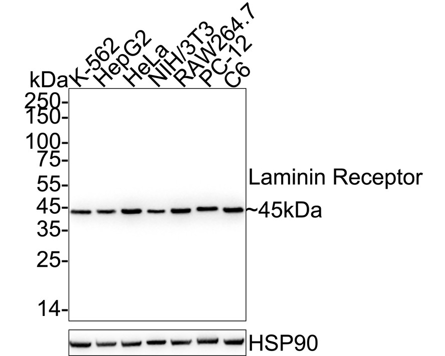
Western blot analysis of 67kDa Laminin Receptor on different lysates with Rabbit anti-67kDa Laminin Receptor antibody at 1/1,000 dilution. Lane 1: K-562 cell lysate, Lane 2: HepG2 cell lysate, Lane 3: HeLa cell lysate, Lane 4: NIH/3T3 cell lysate, Lane 5: RAW264.7 cell lysate, Lane 6: PC-12 cell lysate, Lane 7: C6 cell lysate, Lysates/proteins at 20 µg/Lane. Exposure time: 10 seconds; 4-20% SDS-PAGE gel. Proteins were transferred to a PVDF membrane and blocked with 5% NFDM/TBST for 1 hour at room temperature. The primary antibody at 1/1,000 dilution was used in 5% NFDM/TBST at 4℃ overnight. Goat Anti-Rabbit IgG - HRP Secondary Antibody at 1/50,000 dilution was used for 1 hour at room temperature.ICC/IF
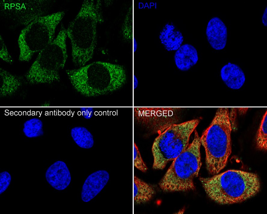
Immunocytochemistry analysis of HeLa cells labeling 67kDa Laminin Receptor with Rabbit anti-67kDa Laminin Receptor antibody at 1/250 dilution. Cells were fixed in 4% paraformaldehyde for 15 minutes at room temperature, permeabilized with 0.1% Triton X-100 in PBS for 15 minutes at room temperature, then blocked with 1% BSA in 10% negative goat serum for 1 hour at room temperature. Cells were then incubated with Rabbit anti-67kDa Laminin Receptor antibody at 1/250 dilution in 1% BSA in PBST overnight at 4 ℃. Goat Anti-Rabbit IgG H&L (488) was used as the secondary antibody at 1/1,000 dilution. PBS instead of the primary antibody was used as the secondary antibody only control. Nuclear DNA was labelled in blue with DAPI. Beta tubulin (red) was stained at 1/100 dilution overnight at +4℃. Goat Anti-Mouse IgG H&L (594) was used as the secondary antibody at 1/1,000 dilution.IHC
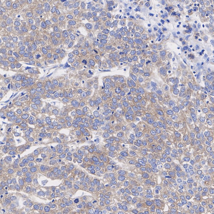
Immunohistochemical analysis of paraffin-embedded human stomach cancer tissue with Rabbit anti-67kDa Laminin Receptor antibody at 1/200 dilution. The section was pre-treated using heat mediated antigen retrieval with Tris-EDTA buffer (pH 9.0) for 20 minutes. The tissues were blocked in 1% BSA for 20 minutes at room temperature, washed with ddH2O and PBS, and then probed with the primary antibody at 1/200 dilution for 1 hour at room temperature. The detection was performed using an HRP conjugated compact polymer system. DAB was used as the chromogen. Tissues were counterstained with hematoxylin and mounted with DPX.FC
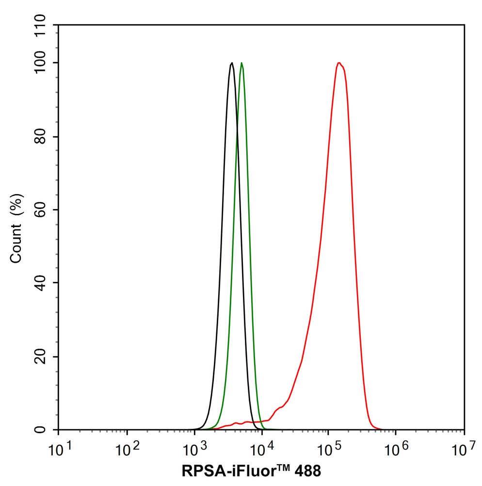
Flow cytometric analysis of HeLa cells labeling 67kDa Laminin Receptor. Cells were fixed and permeabilized. Then stained with the primary antibody (1/1,000) (red) compared with Rabbit IgG Isotype Control (green). After incubation of the primary antibody at +4℃ for an hour, the cells were stained with a iFluor™ 488 conjugate-Goat anti-Rabbit IgG Secondary antibody at 1/1,000 dilution for 30 minutes at +4℃. Unlabelled sample was used as a control (cells without incubation with primary antibody; black).| Product Name | 67kDa Laminin Receptor Recombinant Rabbit Monoclonal Antibody |
|---|---|
| Antibody Type | Primary Antibodies |
| Product description | Laminin receptor (Laminin-R) has a heterodimeric structure similar to that of receptors for other extracellular matrix proteins such as Fibronectin and Vitronectin. Incorporation of Laminin-R into lysosomal membranes makes it possible for lysosomes to attach to surfaces coated with Laminin. This and other properties identify Laminin-R as a member of the integrin family of cell adhesion receptors. The Laminin-R precursor is a polypeptide whose expression is consistently upregulated in aggressive carcinoma. The precursor, which is also identified as p40 ribosome-associated protein, appears to be a multifunctional protein involved in the translational machinery. Laminin-R (also known as colon carcinoma laminin-binding protein) and is found at nine-fold higher levels in colon carcinoma than in adjacent normal colonic epithelium. Additionally, the level of the Laminin-R is higher in the lung cancer cell line than in the lung cell line. |
| Immunogen | Synthetic peptide within Human 67kDa Laminin Receptor aa:250-295 / 295. |
| Clonality | monoclonal |
|---|---|
| Isotype | IgG |
| Host Species | Recombinant rabbit |
| Tested Applications | FCICC/IFIHCWB |
| WB:1:1000-1:2000 ICC/IF:1:250 IHC:1:200-1:1000 FC:1:1000 |
|
| Species Reactivity | HumanMouseRat |
| Concentration | 1mg/ml |
| Purification | Protein A |
| Gene Symbol | RPSA |
|---|---|
| Gene Synonyms | SA LBP LRP p40 uS2 67LR ICAS lamR 37LRP LAMBR LAMR1 LRP/LR LBP/p40 NEM/1CHD4 |
| Gene Full Name | ribosomal protein SA |
| Gene Summary | Laminins, a family of extracellular matrix glycoproteins, are the major noncollagenous constituent of basement membranes. They have been implicated in a wide variety of biological processes including cell adhesion, differentiation, migration, signaling, neurite outgrowth and metastasis. Many of the effects of laminin are mediated through interactions with cell surface receptors. These receptors include members of the integrin family, as well as non-integrin laminin-binding proteins. This gene encodes a high-affinity, non-integrin family, laminin receptor 1. This receptor has been variously called 67 kD laminin receptor, 37 kD laminin receptor precursor (37LRP) and p40 ribosome-associated protein. The amino acid sequence of laminin receptor 1 is highly conserved through evolution, suggesting a key biological function. It has been observed that the level of the laminin receptor transcript is higher in colon carcinoma tissue and lung cancer cell line than their normal counterparts. Also, there is a correlation between the upregulation of this polypeptide in cancer cells and their invasive and metastatic phenotype. Multiple copies of this gene exist, however, most of them are pseudogenes thought to have arisen from retropositional events. Two alternatively spliced transcript variants encoding the same protein have been found for this gene. [provided by RefSeq, Jul 2008] |
| Alternative Names | 34/67 kDa laminin receptor antibody 37 kDa laminin receptor precursor antibody 37/67 kDa laminin receptor antibody 37LRP antibody 40S ribosomal protein SA antibody 67 kDa laminin receptor antibody 67LR antibody Colon carcinoma laminin binding protein antibody Colon carcinoma laminin-binding protein antibody LAMBR antibody Laminin receptor 1 antibody Laminin-binding protein precursor p40 antibody LAMR 1 antibody LamR antibody LAMR1 antibody LBP antibody LBP/p40 antibody LRP antibody LRP/LR antibody Multidrug resistance associated protein MGr1 Ag antibody Multidrug resistance associated protein MGr1Ag antibody Multidrug resistance-associated protein MGr1-Ag antibody NEM/1CHD4 antibody p40 antibody Ribosomal Protein SA antibody rpsA antibody RSSA_HUMAN antibody SA antibody |
| Molecular Weight(MW) | 45kDa |
| Cellular Localization | Cell membrane, Nucleus, Cytoplasm. |
WB
Western blot analysis of 67kDa Laminin Receptor on different lysates with Rabbit anti-67kDa Laminin Receptor antibody at 1/1,000 dilution. Lane 1: K-562 cell lysate, Lane 2: HepG2 cell lysate, Lane 3: HeLa cell lysate, Lane 4: NIH/3T3 cell lysate, Lane 5: RAW264.7 cell lysate, Lane 6: PC-12 cell lysate, Lane 7: C6 cell lysate, Lysates/proteins at 20 µg/Lane. Exposure time: 10 seconds; 4-20% SDS-PAGE gel. Proteins were transferred to a PVDF membrane and blocked with 5% NFDM/TBST for 1 hour at room temperature. The primary antibody at 1/1,000 dilution was used in 5% NFDM/TBST at 4℃ overnight. Goat Anti-Rabbit IgG - HRP Secondary Antibody at 1/50,000 dilution was used for 1 hour at room temperature.
ICC/IF
Immunocytochemistry analysis of HeLa cells labeling 67kDa Laminin Receptor with Rabbit anti-67kDa Laminin Receptor antibody at 1/250 dilution. Cells were fixed in 4% paraformaldehyde for 15 minutes at room temperature, permeabilized with 0.1% Triton X-100 in PBS for 15 minutes at room temperature, then blocked with 1% BSA in 10% negative goat serum for 1 hour at room temperature. Cells were then incubated with Rabbit anti-67kDa Laminin Receptor antibody at 1/250 dilution in 1% BSA in PBST overnight at 4 ℃. Goat Anti-Rabbit IgG H&L (488) was used as the secondary antibody at 1/1,000 dilution. PBS instead of the primary antibody was used as the secondary antibody only control. Nuclear DNA was labelled in blue with DAPI. Beta tubulin (red) was stained at 1/100 dilution overnight at +4℃. Goat Anti-Mouse IgG H&L (594) was used as the secondary antibody at 1/1,000 dilution.
IHC
Immunohistochemical analysis of paraffin-embedded human stomach cancer tissue with Rabbit anti-67kDa Laminin Receptor antibody at 1/200 dilution. The section was pre-treated using heat mediated antigen retrieval with Tris-EDTA buffer (pH 9.0) for 20 minutes. The tissues were blocked in 1% BSA for 20 minutes at room temperature, washed with ddH2O and PBS, and then probed with the primary antibody at 1/200 dilution for 1 hour at room temperature. The detection was performed using an HRP conjugated compact polymer system. DAB was used as the chromogen. Tissues were counterstained with hematoxylin and mounted with DPX.
FC
Flow cytometric analysis of HeLa cells labeling 67kDa Laminin Receptor. Cells were fixed and permeabilized. Then stained with the primary antibody (1/1,000) (red) compared with Rabbit IgG Isotype Control (green). After incubation of the primary antibody at +4℃ for an hour, the cells were stained with a iFluor™ 488 conjugate-Goat anti-Rabbit IgG Secondary antibody at 1/1,000 dilution for 30 minutes at +4℃. Unlabelled sample was used as a control (cells without incubation with primary antibody; black).| Positive Control | K562, A431, RH-35, MCF-7, Hela, HepG2, human gastric carcinoma tissue, mouse kidney tissue, mouse brain tissue. |
|---|---|
| Application Notes | WB:1:1000-1:2000 ICC/IF:1:250 IHC:1:200-1:1000 FC:1:1000 |
| Form | Liquid |
|---|---|
| Storage Instructions | Store at +4℃ after thawing. Aliquot store at -20℃ or -80℃. Avoid repeated freeze / thaw cycles. |
| Storage Buffer | 1*TBS (pH7.4), 1%BSA, 40%Glycerol. Preservative: 0.05% Sodium Azide. |


