WB
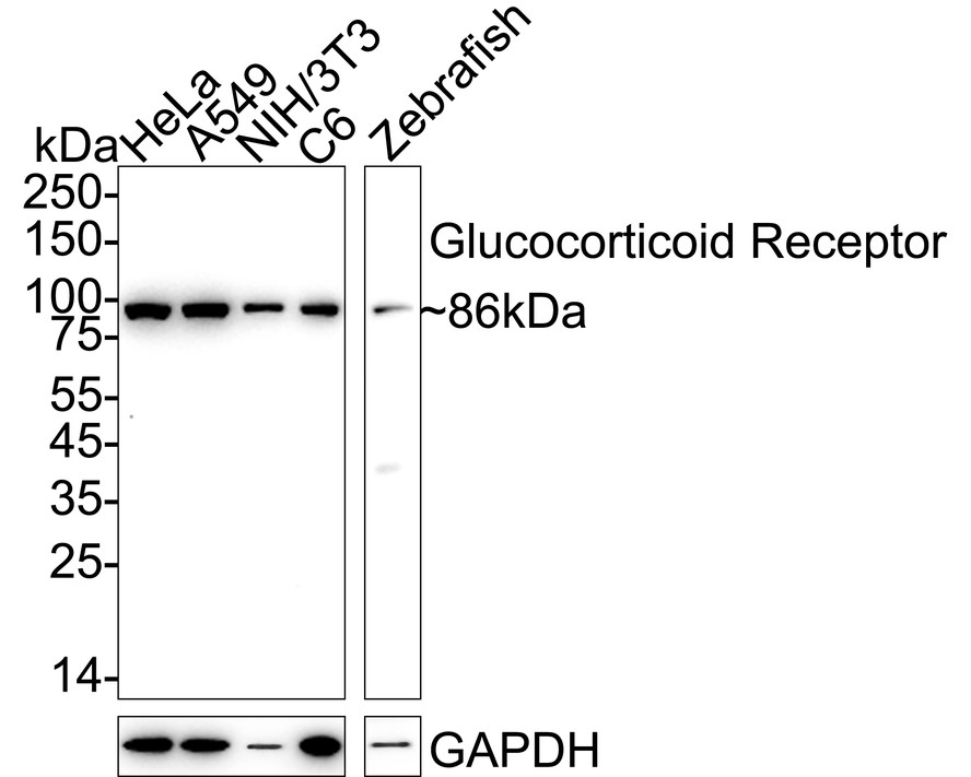
Western blot analysis of Glucocorticoid Receptor on different lysates with Rabbit anti-Glucocorticoid Receptor antibody at 1/2,000 dilution. Lane 1: HeLa cell lysate, Lane 2: A549 cell lysate, Lane 3: NIH/3T3 cell lysate, Lane 4: C6 cell lysate, Lane 5: Zebrafish tissue lysate, Lysates/proteins at 20 µg/Lane. Exposure time: 20 seconds; 4-20% SDS-PAGE gel. Proteins were transferred to a PVDF membrane and blocked with 5% NFDM/TBST for 1 hour at room temperature. The primary antibody at 1/2,000 dilution was used in 5% NFDM/TBST at 4℃ overnight. Goat Anti-Rabbit IgG - HRP Secondary Antibody at 1/50,000 dilution was used for 1 hour at room temperature.ICC/IF
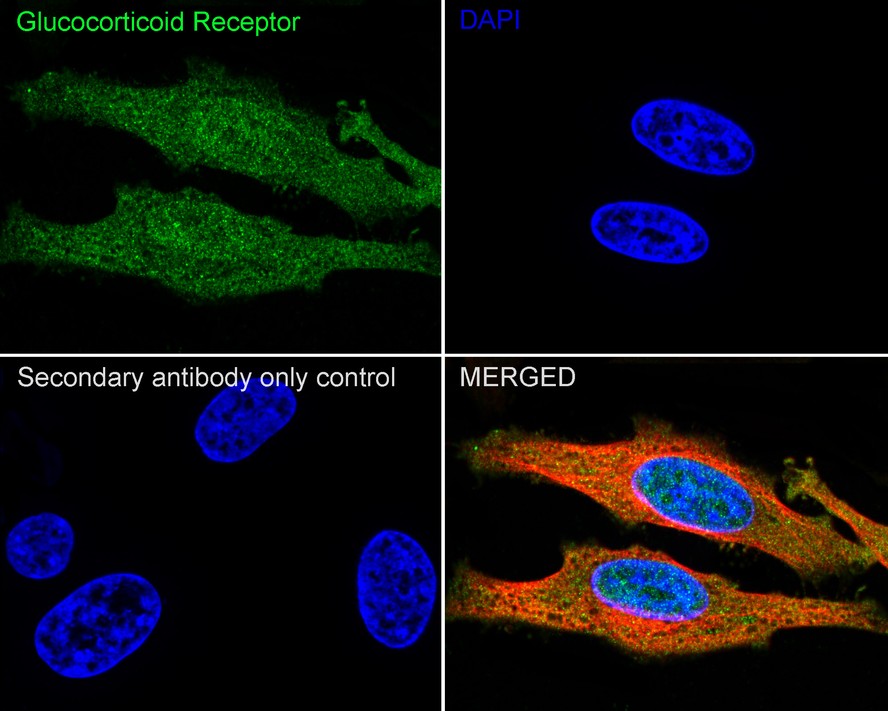
Immunocytochemistry analysis of HeLa cells labeling Glucocorticoid Receptor with Rabbit anti-Glucocorticoid Receptor antibody at 1/100 dilution. Cells were fixed in 4% paraformaldehyde for 15 minutes at room temperature, permeabilized with 0.1% Triton X-100 in PBS for 15 minutes at room temperature, then blocked with 1% BSA in 10% negative goat serum for 1 hour at room temperature. Cells were then incubated with Rabbit anti-Glucocorticoid Receptor antibody at 1/100 dilution in 1% BSA in PBST overnight at 4 ℃. Goat Anti-Rabbit IgG H&L (488) was used as the secondary antibody at 1/1,000 dilution. PBS instead of the primary antibody was used as the secondary antibody only control. Nuclear DNA was labelled in blue with DAPI. Beta tubulin (red) was stained at 1/100 dilution overnight at +4℃. Goat Anti-Mouse IgG H&L (594) was used as the secondary antibody at 1/1,000 dilution.ICC/IF
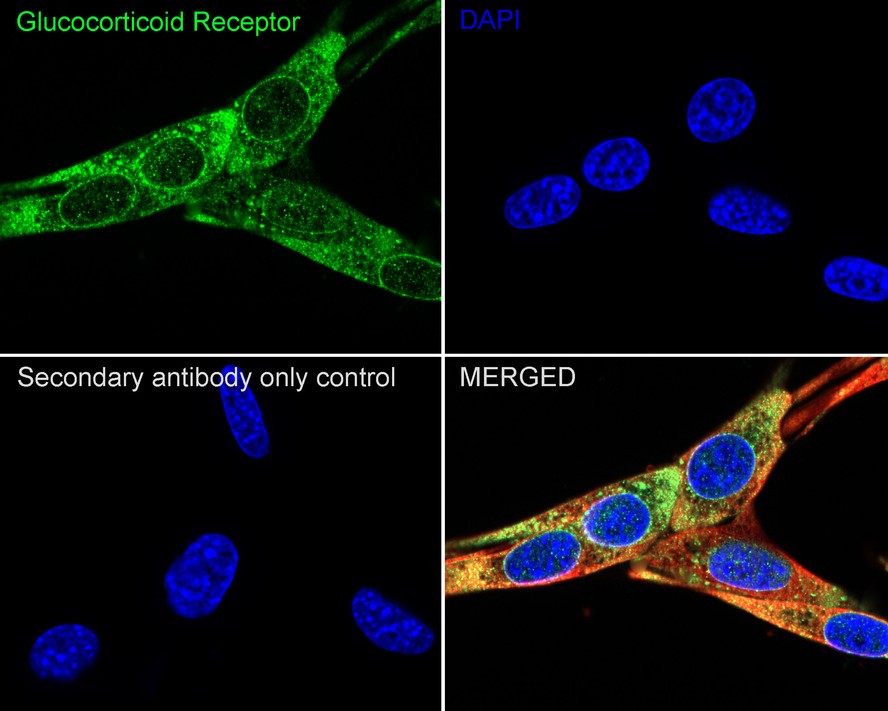
Immunocytochemistry analysis of NIH/3T3 cells labeling Glucocorticoid Receptor with Rabbit anti-Glucocorticoid Receptor antibody at 1/100 dilution. Cells were fixed in 4% paraformaldehyde for 15 minutes at room temperature, permeabilized with 0.1% Triton X-100 in PBS for 15 minutes at room temperature, then blocked with 1% BSA in 10% negative goat serum for 1 hour at room temperature. Cells were then incubated with Rabbit anti-Glucocorticoid Receptor antibody at 1/100 dilution in 1% BSA in PBST overnight at 4 ℃. Goat Anti-Rabbit IgG H&L (488) was used as the secondary antibody at 1/1,000 dilution. PBS instead of the primary antibody was used as the secondary antibody only control. Nuclear DNA was labelled in blue with DAPI. Beta tubulin (red) was stained at 1/100 dilution overnight at +4℃. Goat Anti-Mouse IgG H&L (594) was used as the secondary antibody at 1/1,000 dilution.ICC/IF
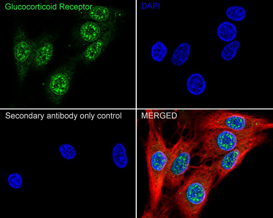
Immunocytochemistry analysis of C6 cells labeling Glucocorticoid Receptor with Rabbit anti-Glucocorticoid Receptor antibody at 1/100 dilution. Cells were fixed in 4% paraformaldehyde for 15 minutes at room temperature, permeabilized with 0.1% Triton X-100 in PBS for 15 minutes at room temperature, then blocked with 1% BSA in 10% negative goat serum for 1 hour at room temperature. Cells were then incubated with Rabbit anti-Glucocorticoid Receptor antibody at 1/100 dilution in 1% BSA in PBST overnight at 4 ℃. Goat Anti-Rabbit IgG H&L (488) was used as the secondary antibody at 1/1,000 dilution. PBS instead of the primary antibody was used as the secondary antibody only control. Nuclear DNA was labelled in blue with DAPI. Beta tubulin (red) was stained at 1/100 dilution overnight at +4℃. Goat Anti-Mouse IgG H&L (594) was used as the secondary antibody at 1/1,000 dilution.FC
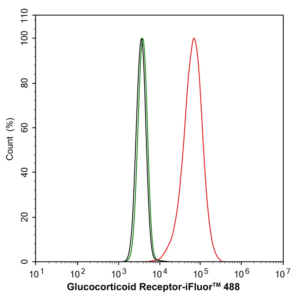
Flow cytometric analysis of HeLa cells labeling Glucocorticoid Receptor. Cells were fixed and permeabilized. Then stained with the primary antibody (1/1,000) (red) compared with Rabbit IgG Isotype Control (green). After incubation of the primary antibody at +4℃ for an hour, the cells were stained with a iFluor™ 488 conjugate-Goat anti-Rabbit IgG Secondary antibody at 1/1,000 dilution for 30 minutes at +4℃. Unlabelled sample was used as a control (cells without incubation with primary antibody; black).| Product Name | Glucocorticoid Receptor Recombinant Rabbit Monoclonal Antibody |
|---|---|
| Antibody Type | Primary Antibodies |
| Product description | The glucocorticoid receptor (GR) is a ubiquitously expressed transcription factor that mediates the effects of glucocorticoids. The most abundant isoform is GR α. GR induces or represses the expression of genes in response to glucocorticoids, mediating such processes as apoptosis, cell growth and differentiation. A significant class of genes suppressed by GR is controlled by the transcription factor AP-1. GR has also been shown to be the limiting factor in the induction of gene expression by glucocorticoids. It has been revealed that GR forms a complex with HSP 90, rendering the non-ligand bound receptor transcriptionally inactive. More importantly, mutant GRs lacking the signaling domain remain constitutively active. |
| Immunogen | Recombinant protein within Human Glucocorticoid Receptor aa 2-220 / 777. |
| Clonality | Monoclonal |
|---|---|
| Isotype | IgG |
| Host Species | Recombinant rabbit |
| Tested Applications | FCICC/IFWB |
| WB:1:2000 ICC/IF:1:100 FC:1:1000 |
|
| Species Reactivity | HumanMouseRatZebrafish |
| Concentration | 1mg/ml |
| Purification | Protein A |
| Alternative Names | GCCR antibody
GCR antibody
GCR_HUMAN antibody
GCRST antibody
glucocorticoid nuclear receptor variant 1 antibody
Glucocorticoid receptor antibody
GR antibody
GRL antibody
Grl1 antibody
nr3c1 antibody
Nuclear receptor subfamily 3 group C member 1 antibody
nuclear receptor subfamily 3 group C member 1 (glucocorticoid receptor) antibody |
|---|---|
| Molecular Weight(MW) | 86kDa |
| Cellular Localization | Cytoplasm, Nucleus, Mitochondrion. |
WB
Western blot analysis of Glucocorticoid Receptor on different lysates with Rabbit anti-Glucocorticoid Receptor antibody at 1/2,000 dilution. Lane 1: HeLa cell lysate, Lane 2: A549 cell lysate, Lane 3: NIH/3T3 cell lysate, Lane 4: C6 cell lysate, Lane 5: Zebrafish tissue lysate, Lysates/proteins at 20 µg/Lane. Exposure time: 20 seconds; 4-20% SDS-PAGE gel. Proteins were transferred to a PVDF membrane and blocked with 5% NFDM/TBST for 1 hour at room temperature. The primary antibody at 1/2,000 dilution was used in 5% NFDM/TBST at 4℃ overnight. Goat Anti-Rabbit IgG - HRP Secondary Antibody at 1/50,000 dilution was used for 1 hour at room temperature.
ICC/IF
Immunocytochemistry analysis of HeLa cells labeling Glucocorticoid Receptor with Rabbit anti-Glucocorticoid Receptor antibody at 1/100 dilution. Cells were fixed in 4% paraformaldehyde for 15 minutes at room temperature, permeabilized with 0.1% Triton X-100 in PBS for 15 minutes at room temperature, then blocked with 1% BSA in 10% negative goat serum for 1 hour at room temperature. Cells were then incubated with Rabbit anti-Glucocorticoid Receptor antibody at 1/100 dilution in 1% BSA in PBST overnight at 4 ℃. Goat Anti-Rabbit IgG H&L (488) was used as the secondary antibody at 1/1,000 dilution. PBS instead of the primary antibody was used as the secondary antibody only control. Nuclear DNA was labelled in blue with DAPI. Beta tubulin (red) was stained at 1/100 dilution overnight at +4℃. Goat Anti-Mouse IgG H&L (594) was used as the secondary antibody at 1/1,000 dilution.
ICC/IF
Immunocytochemistry analysis of NIH/3T3 cells labeling Glucocorticoid Receptor with Rabbit anti-Glucocorticoid Receptor antibody at 1/100 dilution. Cells were fixed in 4% paraformaldehyde for 15 minutes at room temperature, permeabilized with 0.1% Triton X-100 in PBS for 15 minutes at room temperature, then blocked with 1% BSA in 10% negative goat serum for 1 hour at room temperature. Cells were then incubated with Rabbit anti-Glucocorticoid Receptor antibody at 1/100 dilution in 1% BSA in PBST overnight at 4 ℃. Goat Anti-Rabbit IgG H&L (488) was used as the secondary antibody at 1/1,000 dilution. PBS instead of the primary antibody was used as the secondary antibody only control. Nuclear DNA was labelled in blue with DAPI. Beta tubulin (red) was stained at 1/100 dilution overnight at +4℃. Goat Anti-Mouse IgG H&L (594) was used as the secondary antibody at 1/1,000 dilution.
ICC/IF
Immunocytochemistry analysis of C6 cells labeling Glucocorticoid Receptor with Rabbit anti-Glucocorticoid Receptor antibody at 1/100 dilution. Cells were fixed in 4% paraformaldehyde for 15 minutes at room temperature, permeabilized with 0.1% Triton X-100 in PBS for 15 minutes at room temperature, then blocked with 1% BSA in 10% negative goat serum for 1 hour at room temperature. Cells were then incubated with Rabbit anti-Glucocorticoid Receptor antibody at 1/100 dilution in 1% BSA in PBST overnight at 4 ℃. Goat Anti-Rabbit IgG H&L (488) was used as the secondary antibody at 1/1,000 dilution. PBS instead of the primary antibody was used as the secondary antibody only control. Nuclear DNA was labelled in blue with DAPI. Beta tubulin (red) was stained at 1/100 dilution overnight at +4℃. Goat Anti-Mouse IgG H&L (594) was used as the secondary antibody at 1/1,000 dilution.
FC
Flow cytometric analysis of HeLa cells labeling Glucocorticoid Receptor. Cells were fixed and permeabilized. Then stained with the primary antibody (1/1,000) (red) compared with Rabbit IgG Isotype Control (green). After incubation of the primary antibody at +4℃ for an hour, the cells were stained with a iFluor™ 488 conjugate-Goat anti-Rabbit IgG Secondary antibody at 1/1,000 dilution for 30 minutes at +4℃. Unlabelled sample was used as a control (cells without incubation with primary antibody; black).| Application Notes | WB:1:2000 ICC/IF:1:100 FC:1:1000 |
|---|
| Form | Liquid |
|---|---|
| Storage Instructions | Store at +4℃ after thawing. Aliquot store at -20℃ or -80℃. Avoid repeated freeze / thaw cycles. |
| Storage Buffer | 1*TBS (pH7.4), 1%BSA, 40%Glycerol. Preservative: 0.05% Sodium Azide. |


