WB
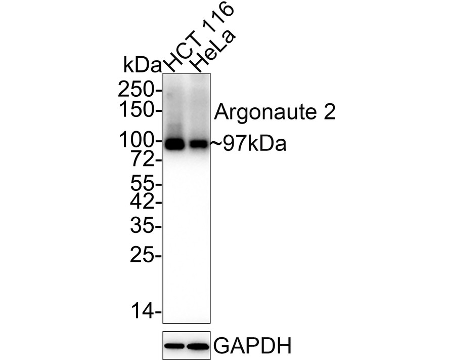
Western blot analysis of Argonaute 2 on different lysates with Rabbit anti-Argonaute 2 antibody at 1/2,000 dilution. Lane 1: HCT 116 cell lysate, Lane 2: HeLa cell lysate, Lysates/proteins at 15 µg/Lane. Exposure time: 1 minute 40 seconds; 4-20% SDS-PAGE gel. Proteins were transferred to a PVDF membrane and blocked with 5% NFDM/TBST for 1 hour at room temperature. The primary antibody at 1/2,000 dilution was used in 5% NFDM/TBST at 4℃ overnight. Goat Anti-Rabbit IgG - HRP Secondary Antibody at 1/50,000 dilution was used for 1 hour at room temperature.ICC/IF
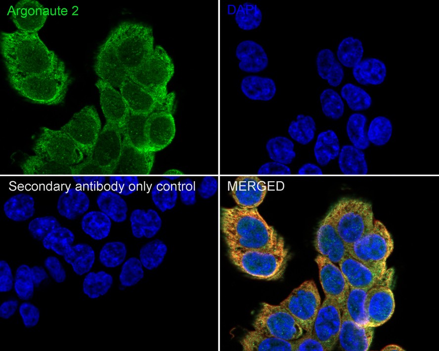
Immunocytochemistry analysis of HCT 116 cells labeling Argonaute 2 with Rabbit anti-Argonaute 2 antibody at 1/500 dilution. Cells were fixed in 4% paraformaldehyde for 20 minutes at room temperature, permeabilized with 0.1% Triton X-100 in PBS for 5 minutes at room temperature, then blocked with 1% BSA in 10% negative goat serum for 1 hour at room temperature. Cells were then incubated with Rabbit anti-Argonaute 2 antibody at 1/500 dilution in 1% BSA in PBST overnight at 4 ℃. Goat Anti-Rabbit IgG H&L (iFluor™ 488) was used as the secondary antibody at 1/1,000 dilution. PBS instead of the primary antibody was used as the secondary antibody only control. Nuclear DNA was labelled in blue with DAPI. Beta tubulin (red) was stained at 1/100 dilution overnight at +4℃. Goat Anti-Mouse IgG H&L (iFluor™ 594) was used as the secondary antibody at 1/1,000 dilution.ICC/IF
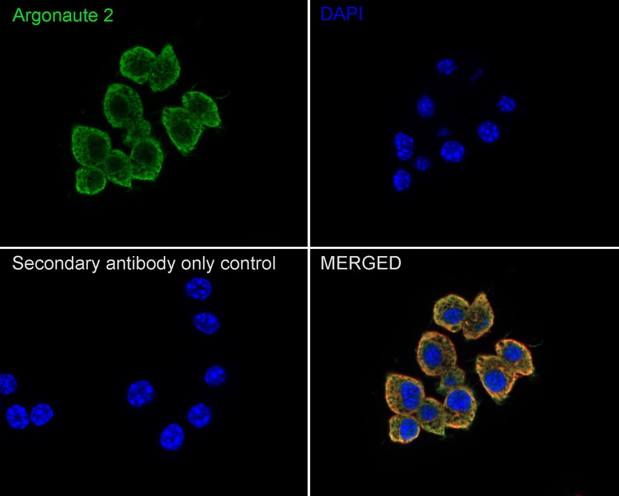
Immunocytochemistry analysis of RAW264.7 cells labeling Argonaute 2 with Rabbit anti-Argonaute 2 antibody at 1/500 dilution. Cells were fixed in 4% paraformaldehyde for 20 minutes at room temperature, permeabilized with 0.1% Triton X-100 in PBS for 5 minutes at room temperature, then blocked with 1% BSA in 10% negative goat serum for 1 hour at room temperature. Cells were then incubated with Rabbit anti-Argonaute 2 antibody at 1/500 dilution in 1% BSA in PBST overnight at 4 ℃. Goat Anti-Rabbit IgG H&L (iFluor™ 488) was used as the secondary antibody at 1/1,000 dilution. PBS instead of the primary antibody was used as the secondary antibody only control. Nuclear DNA was labelled in blue with DAPI. Beta tubulin (red) was stained at 1/100 dilution overnight at +4℃. Goat Anti-Mouse IgG H&L (iFluor™ 594) was used as the secondary antibody at 1/1,000 dilution.ICC/IF
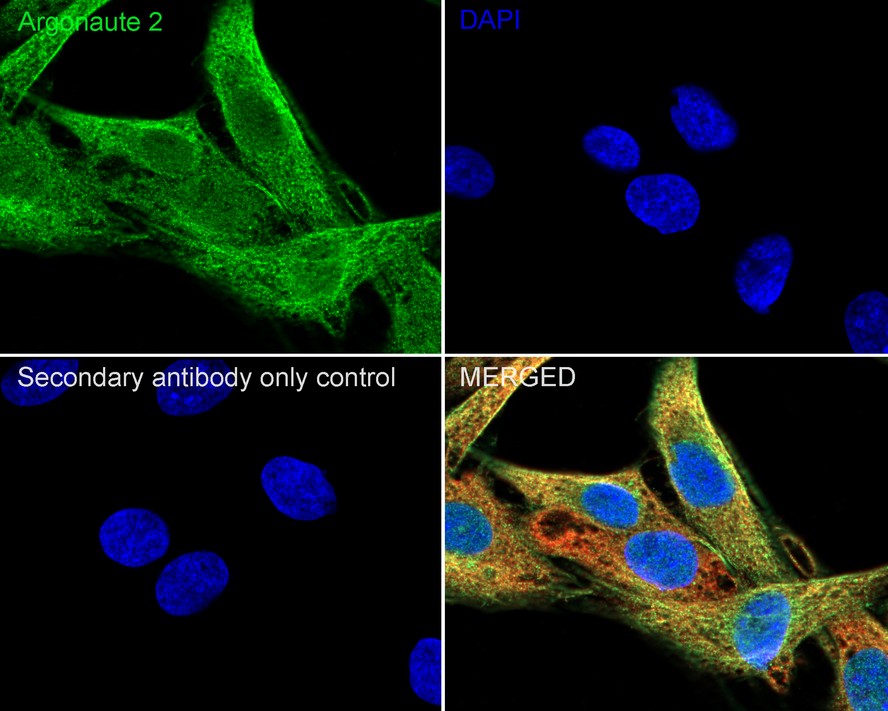
Immunocytochemistry analysis of C6 cells labeling Argonaute 2 with Rabbit anti-Argonaute 2 antibody at 1/500 dilution. Cells were fixed in 4% paraformaldehyde for 20 minutes at room temperature, permeabilized with 0.1% Triton X-100 in PBS for 5 minutes at room temperature, then blocked with 1% BSA in 10% negative goat serum for 1 hour at room temperature. Cells were then incubated with Rabbit anti-Argonaute 2 antibody at 1/500 dilution in 1% BSA in PBST overnight at 4 ℃. Goat Anti-Rabbit IgG H&L (iFluor™ 488) was used as the secondary antibody at 1/1,000 dilution. PBS instead of the primary antibody was used as the secondary antibody only control. Nuclear DNA was labelled in blue with DAPI. Beta tubulin (red) was stained at 1/100 dilution overnight at +4℃. Goat Anti-Mouse IgG H&L (iFluor™ 594) was used as the secondary antibody at 1/1,000 dilution.IHC
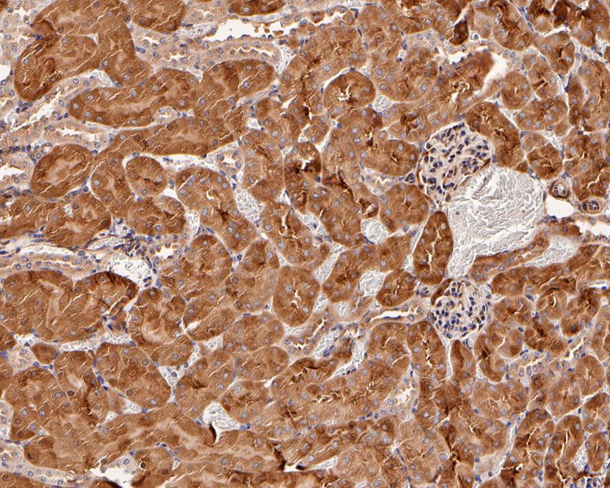
Immunohistochemical analysis of paraffin-embedded mouse kidney tissue with Rabbit anti-Argonaute 2 antibody at 1/4,000 dilution. The section was pre-treated using heat mediated antigen retrieval with Tris-EDTA buffer (pH 9.0) for 20 minutes. The tissues were blocked in 1% BSA for 20 minutes at room temperature, washed with ddH2O and PBS, and then probed with the primary antibody at 1/4,000 dilution for 1 hour at room temperature. The detection was performed using an HRP conjugated compact polymer system. DAB was used as the chromogen. Tissues were counterstained with hematoxylin and mounted with DPX.IHC
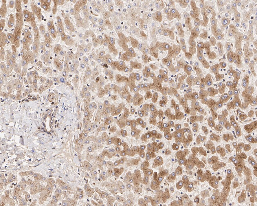
Immunohistochemical analysis of paraffin-embedded human liver tissue with Rabbit anti-Argonaute 2 antibody at 1/1,000 dilution. The section was pre-treated using heat mediated antigen retrieval with sodium citrate buffer (pH 6.0) for 2 minutes. The tissues were blocked in 1% BSA for 20 minutes at room temperature, washed with ddH2O and PBS, and then probed with the primary antibody at 1/1,000 dilution for 1 hour at room temperature. The detection was performed using an HRP conjugated compact polymer system. DAB was used as the chromogen. Tissues were counterstained with hematoxylin and mounted with DPX.FC
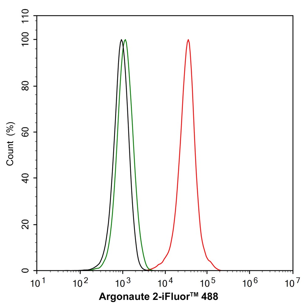
Flow cytometric analysis of RAW264.7 cells labeling Argonaute 2. Cells were fixed and permeabilized. Then stained with the primary antibody (1/1,000) (red) compared with Rabbit IgG Isotype Control (green). After incubation of the primary antibody at +4℃ for an hour, the cells were stained with a iFluor™ 488 conjugate-Goat anti-Rabbit IgG Secondary antibody at 1/1,000 dilution for 30 minutes at +4℃. Unlabelled sample was used as a control (cells without incubation with primary antibody; black).| Product Name | Argonaute 2 Recombinant Rabbit Monoclonal Antibody |
|---|---|
| Antibody Type | Primary Antibodies |
| Product description | Eukaryotic translation initiation factor 2C (eIF2C) proteins (argonaute family) influence RNA interference (RNAi) as components of the RNA-inducible silencing complex (RISC) or microRNA (miRNA)-containing ribonucleoprotein particle (miRNP). Small RNAs, including small interfering RNAs (siRNAs) and miRNAs, can silence target genes through mechanisms that utilize RISC or miRNP particles. eIF2C1 (argonaute 1, AGO1, eIF2C, GERP95, Q99) and Dicer1 play a coordinated role in siRNA-mediated gene silencing. eIF2C2 (Slicer, argonaute 2, AGO2, Q10) is a RISC component that can concentrate in cytoplasmic processing bodies (P-bodies) and catalyze mRNA cleavage. Mammalian P-bodies contain mRNAs and have an association with miRNA-induced translational silencing and siRNA-induced mRNA degradation. Additional eIF2C proteins include eIF2C3 (argonaute 3, AGO3), eIF2C4 (argonaute 4, AGO4) and meIF2c5 (mouse argonaute 5). |
| Immunogen | Synthetic peptide within Human Argonaute 2 aa 361-404 / 859. |
| Clonality | monoclonal |
|---|---|
| Isotype | IgG |
| Host Species | Recombinant rabbit |
| Tested Applications | FCICC/IFIHCWB |
| WB:1:2000 ICC:1:50-1:200 IHC:1:1000-1:4000 FC:1:50-1:100 |
|
| Species Reactivity | HumanMouseRat |
| Concentration | 1mg/ml |
| Purification | Protein A |
| Gene Symbol | AGO2 |
|---|---|
| Gene Synonyms | PPD Q10 CASC7 EIF2C2 LESKRES LINC00980 |
| Gene Full Name | argonaute RISC catalytic component 2 |
| Alternative Names | Ago 2 antibody
AGO2_HUMAN antibody
Argonaute 2 antibody
argonaute 2 RISC catalytic component antibody Argonaute RISC catalytic component 2 antibody Argonaute2 antibody CTA-204B4.6 antibody dAgo2 antibody eIF 2C 2 antibody eIF-2C 2 antibody eIF2C 2 antibody Eif2c2 antibody Eukaryotic translation initiation factor 2C 2 antibody Eukaryotic translation initiation factor 2C subunit 2 antibody hAgo2 antibody MGC3183 antibody PAZ Piwi domain protein antibody PPD antibody Protein argonaute-2 antibody Protein slicer antibody Q10 antibody Slicer protein antibody |
| Molecular Weight(MW) | 97kDa |
| Cellular Localization | Cytoplasm, Nucleus. |
WB
Western blot analysis of Argonaute 2 on different lysates with Rabbit anti-Argonaute 2 antibody at 1/2,000 dilution. Lane 1: HCT 116 cell lysate, Lane 2: HeLa cell lysate, Lysates/proteins at 15 µg/Lane. Exposure time: 1 minute 40 seconds; 4-20% SDS-PAGE gel. Proteins were transferred to a PVDF membrane and blocked with 5% NFDM/TBST for 1 hour at room temperature. The primary antibody at 1/2,000 dilution was used in 5% NFDM/TBST at 4℃ overnight. Goat Anti-Rabbit IgG - HRP Secondary Antibody at 1/50,000 dilution was used for 1 hour at room temperature.
ICC/IF
Immunocytochemistry analysis of HCT 116 cells labeling Argonaute 2 with Rabbit anti-Argonaute 2 antibody at 1/500 dilution. Cells were fixed in 4% paraformaldehyde for 20 minutes at room temperature, permeabilized with 0.1% Triton X-100 in PBS for 5 minutes at room temperature, then blocked with 1% BSA in 10% negative goat serum for 1 hour at room temperature. Cells were then incubated with Rabbit anti-Argonaute 2 antibody at 1/500 dilution in 1% BSA in PBST overnight at 4 ℃. Goat Anti-Rabbit IgG H&L (iFluor™ 488) was used as the secondary antibody at 1/1,000 dilution. PBS instead of the primary antibody was used as the secondary antibody only control. Nuclear DNA was labelled in blue with DAPI. Beta tubulin (red) was stained at 1/100 dilution overnight at +4℃. Goat Anti-Mouse IgG H&L (iFluor™ 594) was used as the secondary antibody at 1/1,000 dilution.
ICC/IF
Immunocytochemistry analysis of RAW264.7 cells labeling Argonaute 2 with Rabbit anti-Argonaute 2 antibody at 1/500 dilution. Cells were fixed in 4% paraformaldehyde for 20 minutes at room temperature, permeabilized with 0.1% Triton X-100 in PBS for 5 minutes at room temperature, then blocked with 1% BSA in 10% negative goat serum for 1 hour at room temperature. Cells were then incubated with Rabbit anti-Argonaute 2 antibody at 1/500 dilution in 1% BSA in PBST overnight at 4 ℃. Goat Anti-Rabbit IgG H&L (iFluor™ 488) was used as the secondary antibody at 1/1,000 dilution. PBS instead of the primary antibody was used as the secondary antibody only control. Nuclear DNA was labelled in blue with DAPI. Beta tubulin (red) was stained at 1/100 dilution overnight at +4℃. Goat Anti-Mouse IgG H&L (iFluor™ 594) was used as the secondary antibody at 1/1,000 dilution.
ICC/IF
Immunocytochemistry analysis of C6 cells labeling Argonaute 2 with Rabbit anti-Argonaute 2 antibody at 1/500 dilution. Cells were fixed in 4% paraformaldehyde for 20 minutes at room temperature, permeabilized with 0.1% Triton X-100 in PBS for 5 minutes at room temperature, then blocked with 1% BSA in 10% negative goat serum for 1 hour at room temperature. Cells were then incubated with Rabbit anti-Argonaute 2 antibody at 1/500 dilution in 1% BSA in PBST overnight at 4 ℃. Goat Anti-Rabbit IgG H&L (iFluor™ 488) was used as the secondary antibody at 1/1,000 dilution. PBS instead of the primary antibody was used as the secondary antibody only control. Nuclear DNA was labelled in blue with DAPI. Beta tubulin (red) was stained at 1/100 dilution overnight at +4℃. Goat Anti-Mouse IgG H&L (iFluor™ 594) was used as the secondary antibody at 1/1,000 dilution.
IHC
Immunohistochemical analysis of paraffin-embedded mouse kidney tissue with Rabbit anti-Argonaute 2 antibody at 1/4,000 dilution. The section was pre-treated using heat mediated antigen retrieval with Tris-EDTA buffer (pH 9.0) for 20 minutes. The tissues were blocked in 1% BSA for 20 minutes at room temperature, washed with ddH2O and PBS, and then probed with the primary antibody at 1/4,000 dilution for 1 hour at room temperature. The detection was performed using an HRP conjugated compact polymer system. DAB was used as the chromogen. Tissues were counterstained with hematoxylin and mounted with DPX.
IHC
Immunohistochemical analysis of paraffin-embedded human liver tissue with Rabbit anti-Argonaute 2 antibody at 1/1,000 dilution. The section was pre-treated using heat mediated antigen retrieval with sodium citrate buffer (pH 6.0) for 2 minutes. The tissues were blocked in 1% BSA for 20 minutes at room temperature, washed with ddH2O and PBS, and then probed with the primary antibody at 1/1,000 dilution for 1 hour at room temperature. The detection was performed using an HRP conjugated compact polymer system. DAB was used as the chromogen. Tissues were counterstained with hematoxylin and mounted with DPX.
FC
Flow cytometric analysis of RAW264.7 cells labeling Argonaute 2. Cells were fixed and permeabilized. Then stained with the primary antibody (1/1,000) (red) compared with Rabbit IgG Isotype Control (green). After incubation of the primary antibody at +4℃ for an hour, the cells were stained with a iFluor™ 488 conjugate-Goat anti-Rabbit IgG Secondary antibody at 1/1,000 dilution for 30 minutes at +4℃. Unlabelled sample was used as a control (cells without incubation with primary antibody; black).| Positive Control | AGS, MCF-7, Hela, human kidney tissue, mouse kidney tissue, mouse stomach tissue. |
|---|---|
| Application Notes | WB:1:2000 ICC:1:50-1:200 IHC:1:1000-1:4000 FC:1:50-1:100 |
| Form | Liquid |
|---|---|
| Storage Instructions | Store at +4℃ after thawing. Aliquot store at -20℃ or -80℃. Avoid repeated freeze / thaw cycles. |
| Storage Buffer | 1*TBS (pH7.4), 1%BSA, 40%Glycerol. Preservative: 0.05% Sodium Azide. |


