WB
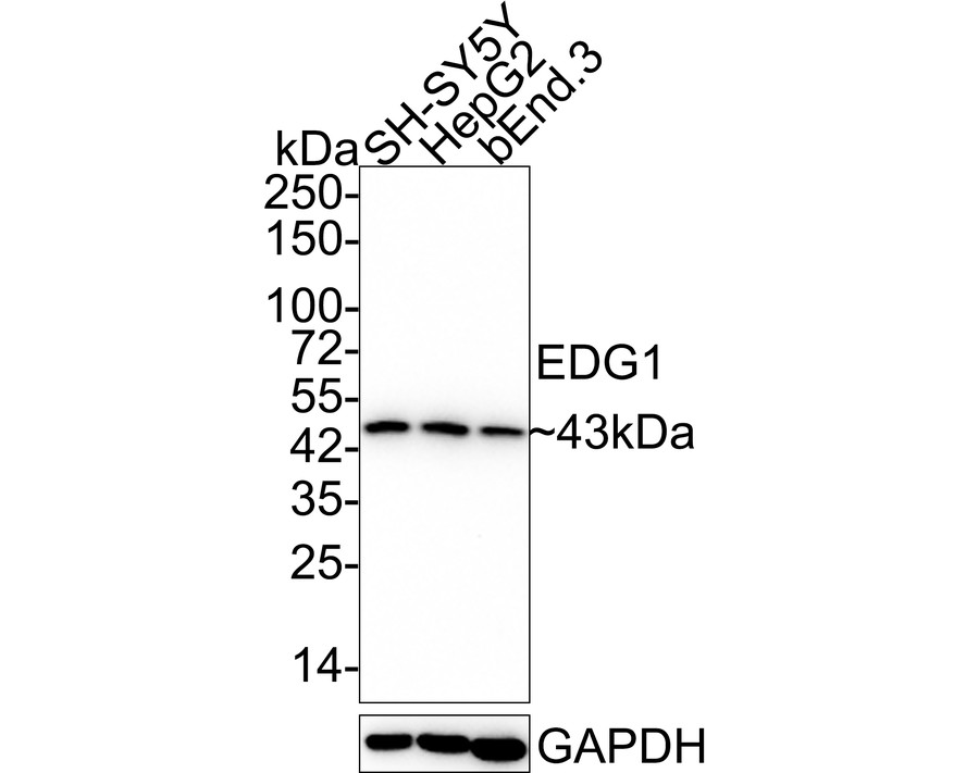
Western blot analysis of EDG1 on different lysates with Rabbit anti-EDG1 antibody at 1/5,000 dilution.Lysates/proteins at 20 µg/Lane.Exposure time: 1 minute 21 seconds;4-20% SDS-PAGE gel.Proteins were transferred to a PVDF membrane and blocked with 5% NFDM/TBST for 1 hour at room temperature. The primary antibody at 1/5,000 dilution was used in 5% NFDM/TBST at 4℃ overnight. Goat Anti-Rabbit IgG - HRP Secondary Antibody at 1:50,000 dilution was used for 1 hour at room temperature.WB
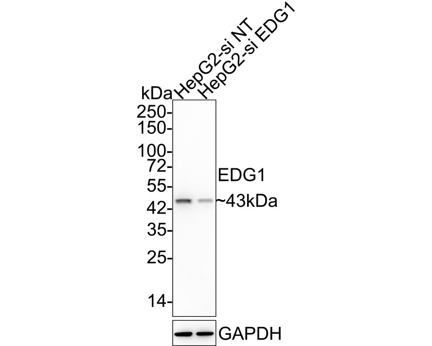
Western blot analysis of EDG1 on different lysates with Rabbit anti-EDG1 antibody at 1/5,000 dilution.Lysates/proteins at 10 µg/Lane.Exposure time: 2 minutes; 4-20% SDS-PAGE gel. Proteins were transferred to a PVDF membrane and blocked with 5% NFDM/TBST for 1 hour at room temperature. The primary antibody at 1/5,000 dilution was used in 5% NFDM/TBST at 4℃ overnight. Goat Anti-Rabbit IgG - HRP Secondary Antibody at 1/50,000 dilution was used for 1 hour at room temperature.WB
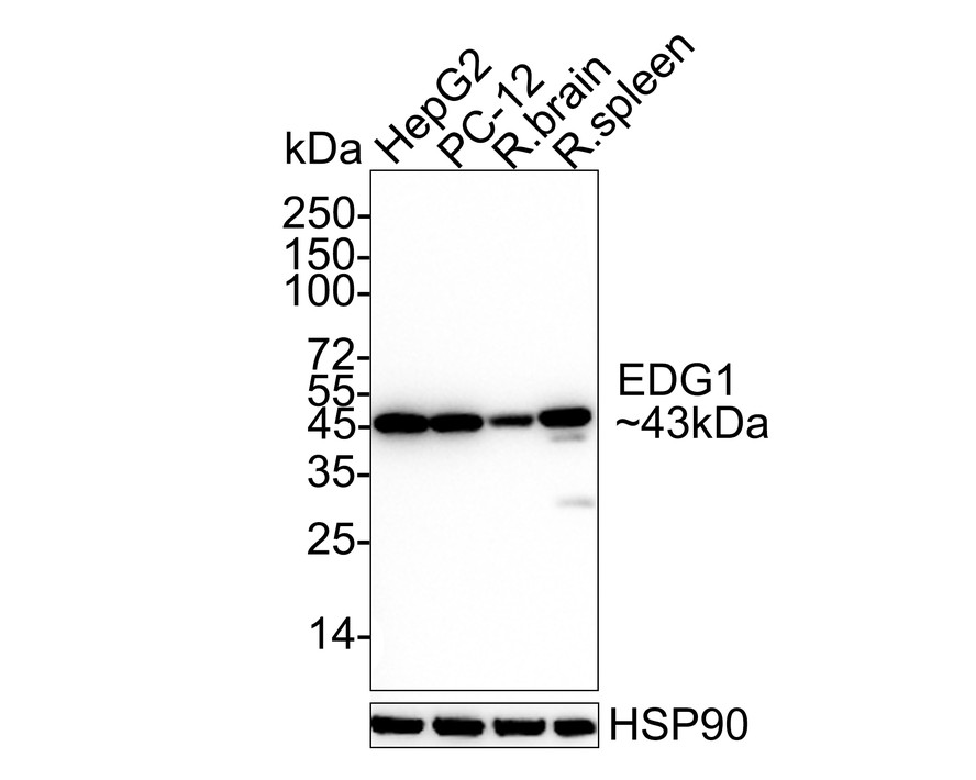
Western blot analysis of EDG1 on different lysates with Rabbit anti-EDG1 antibody at 1/2,000 dilution.Lysates/proteins at 20/40 µg/Lane.Exposure time: 60 seconds;4-20% SDS-PAGE gel. Proteins were transferred to a PVDF membrane and blocked with 5% NFDM/TBST for 1 hour at room temperature. The primary antibody at 1/2,000 dilution was used in 5% NFDM/TBST at 4℃ overnight. Goat Anti-Rabbit IgG - HRP Secondary Antibody at 1/50,000 dilution was used for 1 hour at room temperature.IP
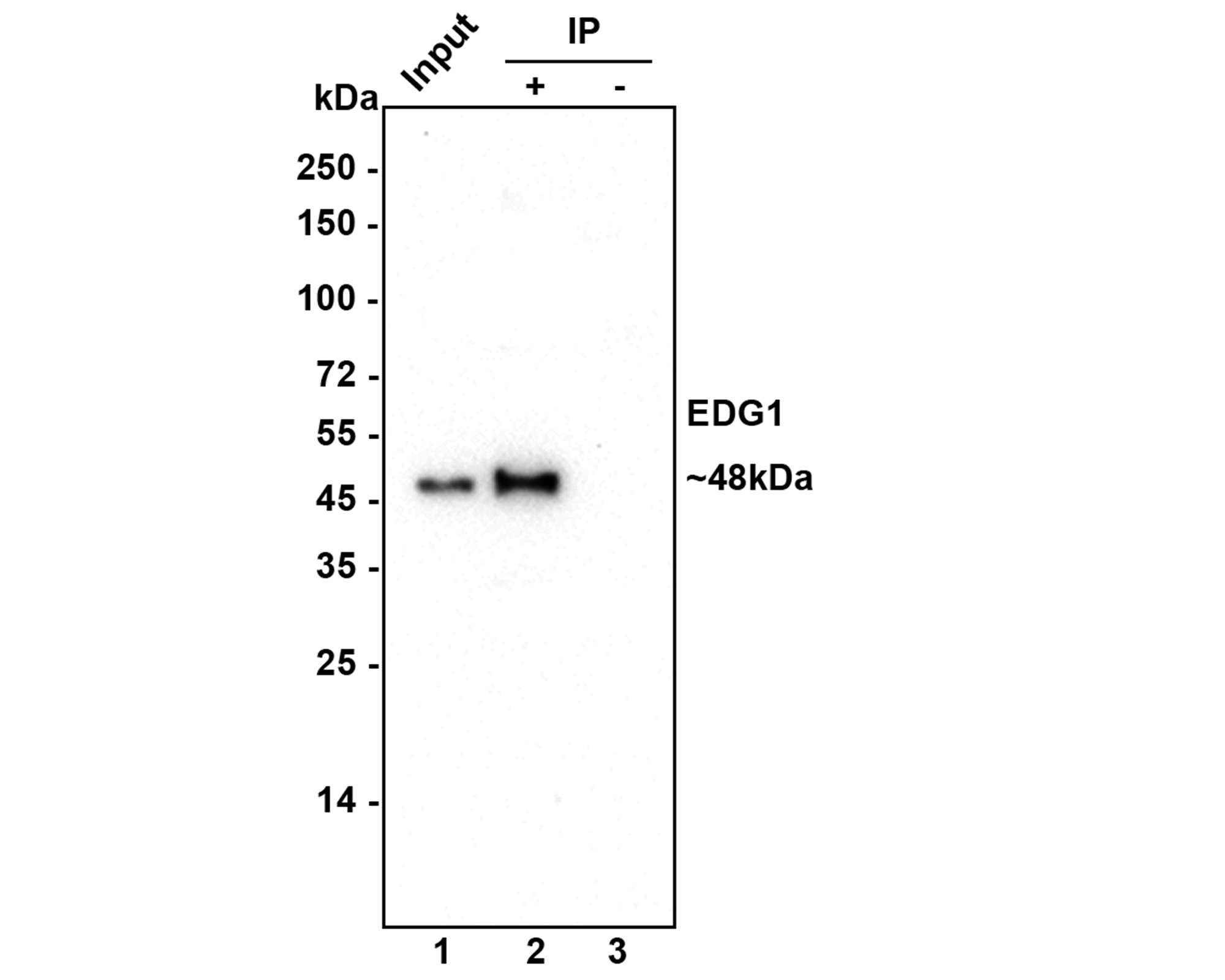
EDG1 was immunoprecipitated in 0.2mg HepG2 cell lysate with Rabbit anti-EDG1 antibody at 2 µg/10 µl beads. Western blot was performed from the immunoprecipitate using Rabbit anti-EDG1 antibody at 1/5,000 dilution. Anti-Rabbit IgG for IP Nano-secondary antibody (NBI01H) at 1/5,000 dilution was used for 1 hour at room temperature.Lane 1: HepG2 cell lysate (input);Lane 2:Rabbit anti-EDG1 antibody IP in HepG2 cell lysate;Lane 3: Rabbit IgG instead of Rabbit anti-EDG1 antibody in HepG2 cell lysate Blocking/Dilution buffer: 5% NFDM/TBST Exposure time: 4 mintues| Product Name | EDG1 Recombinant Rabbit Monoclonal Antibody |
|---|---|
| Antibody Type | Primary Antibodies |
| Immunogen | Synthetic peptide within Human EDG1 aa 2-51 / 382. |
| Clonality | Monoclonal |
|---|---|
| Isotype | IgG |
| Host Species | Recombinant rabbit |
| Tested Applications | IPWB |
| WB:1:2000-1:5000 IP:Use at anassay dependent concentration. |
|
| Species Reactivity | HumanMouseRat |
| Concentration | 1mg/ml |
| Purification | Protein A |
| Gene Symbol | S1PR1 |
|---|---|
| Gene Synonyms | EDG1 S1P1 CD363 ECGF1 EDG-1 CHEDG1 D1S3362 |
| Gene Full Name | sphingosine-1-phosphate receptor 1 |
| Gene Summary | The protein encoded by this gene is structurally similar to G protein-coupled receptors and is highly expressed in endothelial cells. It binds the ligand sphingosine-1-phosphate with high affinity and high specificity, and suggested to be involved in the processes that regulate the differentiation of endothelial cells. Activation of this receptor induces cell-cell adhesion. Alternative splicing results in multiple transcript variants. [provided by RefSeq, Mar 2016] |
| Molecular Weight(MW) | 43 kDa |
| Cellular Localization | Cell membrane. |
WB
Western blot analysis of EDG1 on different lysates with Rabbit anti-EDG1 antibody at 1/5,000 dilution.Lysates/proteins at 20 µg/Lane.Exposure time: 1 minute 21 seconds;4-20% SDS-PAGE gel.Proteins were transferred to a PVDF membrane and blocked with 5% NFDM/TBST for 1 hour at room temperature. The primary antibody at 1/5,000 dilution was used in 5% NFDM/TBST at 4℃ overnight. Goat Anti-Rabbit IgG - HRP Secondary Antibody at 1:50,000 dilution was used for 1 hour at room temperature.
WB
Western blot analysis of EDG1 on different lysates with Rabbit anti-EDG1 antibody at 1/5,000 dilution.Lysates/proteins at 10 µg/Lane.Exposure time: 2 minutes; 4-20% SDS-PAGE gel. Proteins were transferred to a PVDF membrane and blocked with 5% NFDM/TBST for 1 hour at room temperature. The primary antibody at 1/5,000 dilution was used in 5% NFDM/TBST at 4℃ overnight. Goat Anti-Rabbit IgG - HRP Secondary Antibody at 1/50,000 dilution was used for 1 hour at room temperature.
WB
Western blot analysis of EDG1 on different lysates with Rabbit anti-EDG1 antibody at 1/2,000 dilution.Lysates/proteins at 20/40 µg/Lane.Exposure time: 60 seconds;4-20% SDS-PAGE gel. Proteins were transferred to a PVDF membrane and blocked with 5% NFDM/TBST for 1 hour at room temperature. The primary antibody at 1/2,000 dilution was used in 5% NFDM/TBST at 4℃ overnight. Goat Anti-Rabbit IgG - HRP Secondary Antibody at 1/50,000 dilution was used for 1 hour at room temperature.
IP
EDG1 was immunoprecipitated in 0.2mg HepG2 cell lysate with Rabbit anti-EDG1 antibody at 2 µg/10 µl beads. Western blot was performed from the immunoprecipitate using Rabbit anti-EDG1 antibody at 1/5,000 dilution. Anti-Rabbit IgG for IP Nano-secondary antibody (NBI01H) at 1/5,000 dilution was used for 1 hour at room temperature.Lane 1: HepG2 cell lysate (input);Lane 2:Rabbit anti-EDG1 antibody IP in HepG2 cell lysate;Lane 3: Rabbit IgG instead of Rabbit anti-EDG1 antibody in HepG2 cell lysate Blocking/Dilution buffer: 5% NFDM/TBST Exposure time: 4 mintues| Application Notes | WB:1:2000-1:5000 IP:Use at anassay dependent concentration. |
|---|
| Form | Liquid |
|---|---|
| Storage Instructions | Shipped at 4℃. Store at +4℃ short term (1-2 weeks). Store at -20℃ long term. |
| Storage Buffer | TBS (pH7.4), 0.05% BSA, 40% Glycerol. Preservative: 0.05% Sodium Azide. |


