WB
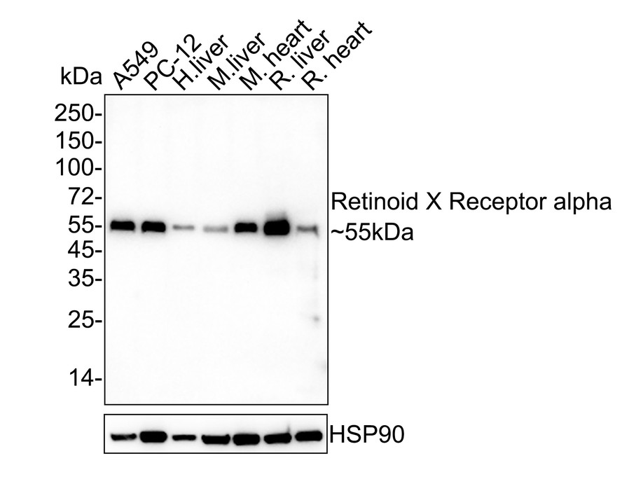
Western blot analysis of Retinoid X Receptor alpha on different lysates with Rabbit anti-Retinoid X Receptor alpha antibody at 1/500 dilution. Lane 1: A549 cell lysate, Lane 2: PC-12 cell lysate, Lane 3: Human liver tissue lysate, Lane 4: Mouse liver tissue lysate, Lane 5: Mouse heart tissue lysate, Lane 6: Rat liver tissue lysate, Lane 7: Rat heart tissue lysate, Cell lysates/proteins at 20 µg/Lane. Tissue lysates/proteins at 40 µg/Lane. Exposure time: 3 minutes; 4-20% SDS-PAGE gel. Proteins were transferred to a PVDF membrane and blocked with 5% NFDM/TBST for 1 hour at room temperature. The primary antibody at 1/500 dilution was used in 5% NFDM/TBST at 4℃ overnight. Goat Anti-Rabbit IgG - HRP Secondary Antibody at 1/50,000 dilution was used for 1 hour at room temperature.IHC
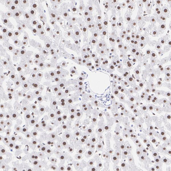
Immunohistochemical analysis of paraffin-embedded rat liver tissue with Rabbit anti-Retinoid X Receptor alpha antibody at 1/1,000 dilution. The section was pre-treated using heat mediated antigen retrieval with Tris-EDTA buffer (pH 9.0) for 20 minutes. The tissues were blocked in 1% BSA for 20 minutes at room temperature, washed with ddH2O and PBS, and then probed with the primary antibody at 1/1,000 dilution for 1 hour at room temperature. The detection was performed using an HRP conjugated compact polymer system. DAB was used as the chromogen. Tissues were counterstained with hematoxylin and mounted with DPX.IHC
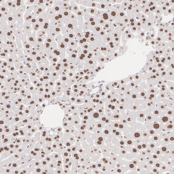
Immunohistochemical analysis of paraffin-embedded mouse liver tissue with Rabbit anti-Retinoid X Receptor alpha antibody at 1/1,000 dilution. The section was pre-treated using heat mediated antigen retrieval with Tris-EDTA buffer (pH 9.0) for 20 minutes. The tissues were blocked in 1% BSA for 20 minutes at room temperature, washed with ddH2O and PBS, and then probed with the primary antibody at 1/1,000 dilution for 1 hour at room temperature. The detection was performed using an HRP conjugated compact polymer system. DAB was used as the chromogen. Tissues were counterstained with hematoxylin and mounted with DPX.IHC
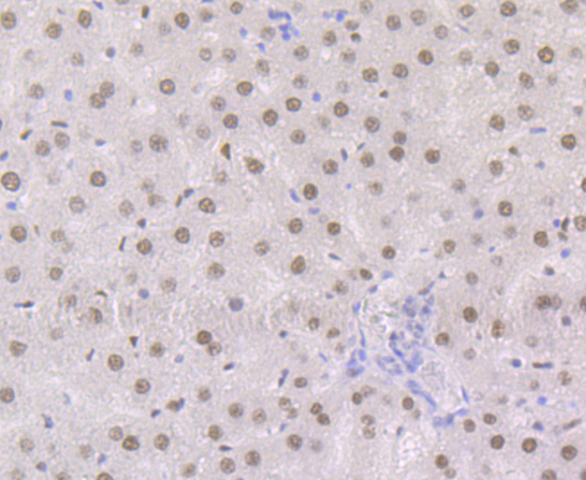
Immunohistochemical analysis of paraffin-embedded rat liver tissue using anti-Retinoid X Receptor alpha antibody. The section was pre-treated using heat mediated antigen retrieval with Tris-EDTA buffer (pH 8.0-8.4) for 20 minutes.The tissues were blocked in 5% BSA for 30 minutes at room temperature, washed with ddH2O and PBS, and then probed with the primary antibody (1/50) for 30 minutes at room temperature. The detection was performed using an HRP conjugated compact polymer system. DAB was used as the chromogen. Tissues were counterstained with hematoxylin and mounted with DPX.IHC
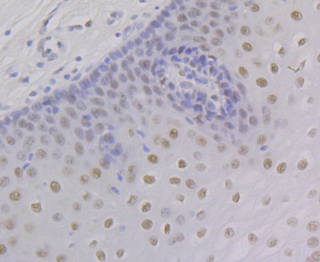
Immunohistochemical analysis of paraffin-embedded human esophagus tissue using anti-Retinoid X Receptor alpha antibody. The section was pre-treated using heat mediated antigen retrieval with Tris-EDTA buffer (pH 8.0-8.4) for 20 minutes.The tissues were blocked in 5% BSA for 30 minutes at room temperature, washed with ddH2O and PBS, and then probed with the primary antibody (1/50) for 30 minutes at room temperature. The detection was performed using an HRP conjugated compact polymer system. DAB was used as the chromogen. Tissues were counterstained with hematoxylin and mounted with DPX.| Product Name | Retinoid X Receptor alpha Recombinant Rabbit Monoclonal Antibody |
|---|---|
| Antibody Type | Primary Antibodies |
| Product description | Receptor for retinoic acid. Retinoic acid receptors bind as heterodimers to their target response elements in response to their ligands, all-trans or 9-cis retinoic acid, and regulate gene expression in various biological processes. The RAR/RXR heterodimers bind to the retinoic acid response elements (RARE) composed of tandem 5'-AGGTCA-3' sites known as DR1-DR5. The high affinity ligand for RXRs is 9-cis retinoic acid. RXRA serves as a common heterodimeric partner for a number of nuclear receptors. In the absence of ligand, the RXR-RAR heterodimers associate with a multiprotein complex containing transcription corepressors that induce histone acetylation, chromatin condensation and transcriptional suppression. On ligand binding, the corepressors dissociate from the receptors and associate with the coactivators leading to transcriptional activation. The RXRA/PPARA heterodimer is required for PPARA transcriptional activity on fatty acid oxidation genes such as ACOX1 and the P450 system genes. |
| Immunogen | Recombinant protein corresponding to Human Retinoid X Receptor alpha aa 1-50 / 462. |
| Clonality | monoclonal |
|---|---|
| Isotype | IgG |
| Host Species | Recombinant rabbit |
| Tested Applications | IHCWB |
| WB:1:500-1:2000 IHC:1:50-1:400 |
|
| Species Reactivity | HumanMouseRat |
| Concentration | 1mg/ml |
| Purification | Protein A |
| Gene Symbol | RXRA |
|---|---|
| Gene Synonyms | NR2B1 RXRalpha RXR-alpha |
| Gene Full Name | retinoid X receptor alpha |
| Gene Summary | Retinoid X receptors (RXRs) and retinoic acid receptors (RARs) are nuclear receptors that mediate the biological effects of retinoids by their involvement in retinoic acid-mediated gene activation. These receptors function as transcription factors by binding as homodimers or heterodimers to specific sequences in the promoters of target genes. The protein encoded by this gene is a member of the steroid and thyroid hormone receptor superfamily of transcriptional regulators. Alternative splicing of this gene results in multiple transcript variants. [provided by RefSeq, May 2014] |
| Alternative Names | FLJ00280 antibody FLJ00318 antibody FLJ16020 antibody FLJ16733 antibody MGC102720 antibody NR2B1 antibody Nuclear receptor subfamily 2 group B member 1 antibody OTTHUMP00000022510 antibody Retinoic acid receptor RXR alpha antibody Retinoic acid receptor RXR-alpha antibody Retinoid X nuclear receptor alpha antibody Retinoid X receptor alpha antibody RXR alpha1 antibody Rxra antibody RXRA_HUMAN antibody RXRalpha1 antibody |
| Molecular Weight(MW) | 51kDa(Observed band size: 55kDa) |
| Cellular Localization | Mitochondrion, Nucleus, Cytoplasm. |
WB
Western blot analysis of Retinoid X Receptor alpha on different lysates with Rabbit anti-Retinoid X Receptor alpha antibody at 1/500 dilution. Lane 1: A549 cell lysate, Lane 2: PC-12 cell lysate, Lane 3: Human liver tissue lysate, Lane 4: Mouse liver tissue lysate, Lane 5: Mouse heart tissue lysate, Lane 6: Rat liver tissue lysate, Lane 7: Rat heart tissue lysate, Cell lysates/proteins at 20 µg/Lane. Tissue lysates/proteins at 40 µg/Lane. Exposure time: 3 minutes; 4-20% SDS-PAGE gel. Proteins were transferred to a PVDF membrane and blocked with 5% NFDM/TBST for 1 hour at room temperature. The primary antibody at 1/500 dilution was used in 5% NFDM/TBST at 4℃ overnight. Goat Anti-Rabbit IgG - HRP Secondary Antibody at 1/50,000 dilution was used for 1 hour at room temperature.
IHC
Immunohistochemical analysis of paraffin-embedded rat liver tissue with Rabbit anti-Retinoid X Receptor alpha antibody at 1/1,000 dilution. The section was pre-treated using heat mediated antigen retrieval with Tris-EDTA buffer (pH 9.0) for 20 minutes. The tissues were blocked in 1% BSA for 20 minutes at room temperature, washed with ddH2O and PBS, and then probed with the primary antibody at 1/1,000 dilution for 1 hour at room temperature. The detection was performed using an HRP conjugated compact polymer system. DAB was used as the chromogen. Tissues were counterstained with hematoxylin and mounted with DPX.
IHC
Immunohistochemical analysis of paraffin-embedded mouse liver tissue with Rabbit anti-Retinoid X Receptor alpha antibody at 1/1,000 dilution. The section was pre-treated using heat mediated antigen retrieval with Tris-EDTA buffer (pH 9.0) for 20 minutes. The tissues were blocked in 1% BSA for 20 minutes at room temperature, washed with ddH2O and PBS, and then probed with the primary antibody at 1/1,000 dilution for 1 hour at room temperature. The detection was performed using an HRP conjugated compact polymer system. DAB was used as the chromogen. Tissues were counterstained with hematoxylin and mounted with DPX.
IHC
Immunohistochemical analysis of paraffin-embedded rat liver tissue using anti-Retinoid X Receptor alpha antibody. The section was pre-treated using heat mediated antigen retrieval with Tris-EDTA buffer (pH 8.0-8.4) for 20 minutes.The tissues were blocked in 5% BSA for 30 minutes at room temperature, washed with ddH2O and PBS, and then probed with the primary antibody (1/50) for 30 minutes at room temperature. The detection was performed using an HRP conjugated compact polymer system. DAB was used as the chromogen. Tissues were counterstained with hematoxylin and mounted with DPX.
IHC
Immunohistochemical analysis of paraffin-embedded human esophagus tissue using anti-Retinoid X Receptor alpha antibody. The section was pre-treated using heat mediated antigen retrieval with Tris-EDTA buffer (pH 8.0-8.4) for 20 minutes.The tissues were blocked in 5% BSA for 30 minutes at room temperature, washed with ddH2O and PBS, and then probed with the primary antibody (1/50) for 30 minutes at room temperature. The detection was performed using an HRP conjugated compact polymer system. DAB was used as the chromogen. Tissues were counterstained with hematoxylin and mounted with DPX.| Positive Control | MCF-7, rat liver tissue, human esophagus tissue. |
|---|---|
| Application Notes | WB:1:500-1:2000 IHC:1:50-1:400 |
| Form | Liquid |
|---|---|
| Storage Instructions | Store at +4℃ after thawing. Aliquot store at -20℃. Avoid repeated freeze / thaw cycles. |
| Storage Buffer | 1*TBS (pH7.4), 1%BSA, 40%Glycerol. Preservative: 0.05% Sodium Azide. |


