Application
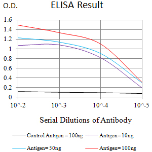
Figure 1:Black line: Control Antigen (100 ng);Purple line: Antigen (10ng); Blue line: Antigen (50 ng); Red line:Antigen (100 ng)Application
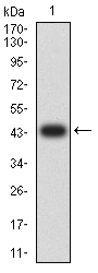
Figure 2:Western blot analysis using ATXN1 mAb against human ATXN1 (AA: 645-815) recombinant protein. (Expected MW is 44.1 kDa)Application
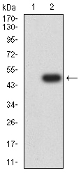
Figure 3:Western blot analysis using ATXN1 mAb against HEK293 (1) and ATXN1 (AA: 645-815)-hIgGFc transfected HEK293 (2) cell lysate.Application
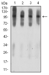
Figure 4:Western blot analysis using ATXN1 mouse mAb against C6 (1), COS7 (2), NIH/3T3 (3), and HL-60 (4) cell lysate.Application
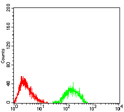
Figure 5:Flow cytometric analysis of Jurkat cells using ATXN1 mouse mAb (green) and negative control (red).Application
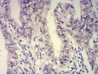
Figure 6:Immunohistochemical analysis of paraffin-embedded endometrial cancer tissues using ATXN1 mouse mAb with DAB staining.| Product Name | ATXN1 |
|---|---|
| Antibody Type | Primary Antibodies |
| Antigen Alias | ATX1; SCA1; D6S504E |
| Immunogen | Purified recombinant fragment of human ATXN1 (AA: 645-815) expressed in E. Coli. |
| Clonality | Monoclonal |
|---|---|
| Host Species | Unknow |
| Tested Applications | WBIHCFCMICC/IF |
| WB:1:250~1:1000 IHC:1:20~1:100 ICC/IF:1:100~1:200 Notes:Optimal dilutions/concentrations should be determined by the researcher. |
|
| Species Reactivity | HumanMouseMonkeyRat |
| Alternative Names | ATX1 SCA1 D6S504E |
|---|---|
| Molecular Weight(MW) | 86.9kDa |
| Entrez Gene | 6310 |
|---|
Application
Figure 1:Black line: Control Antigen (100 ng);Purple line: Antigen (10ng); Blue line: Antigen (50 ng); Red line:Antigen (100 ng)
Application
Figure 2:Western blot analysis using ATXN1 mAb against human ATXN1 (AA: 645-815) recombinant protein. (Expected MW is 44.1 kDa)
Application
Figure 3:Western blot analysis using ATXN1 mAb against HEK293 (1) and ATXN1 (AA: 645-815)-hIgGFc transfected HEK293 (2) cell lysate.
Application
Figure 4:Western blot analysis using ATXN1 mouse mAb against C6 (1), COS7 (2), NIH/3T3 (3), and HL-60 (4) cell lysate.
Application
Figure 5:Flow cytometric analysis of Jurkat cells using ATXN1 mouse mAb (green) and negative control (red).
Application
Figure 6:Immunohistochemical analysis of paraffin-embedded endometrial cancer tissues using ATXN1 mouse mAb with DAB staining.| Application Notes | WB:1:250~1:1000 IHC:1:20~1:100 ICC/IF:1:100~1:200 Notes:Optimal dilutions/concentrations should be determined by the researcher. |
|---|
| Form | Purified antibody in PBS with 0.05% sodium azide |
|---|---|
| Storage Instructions | 4℃; -20℃ for long term storage |


