


WB
Western blot analysis using Histone H3 antibody against PANC-1 (1) cell lysate.15% SDS-PAGE gel.Sample loading: 20μg /lane. Transfer the proteins onto a PVDF membrane (OM790003), and block it with TBST (OM750016) plus skimmed milk powder for one hour. Dilute the primary antibody with the antibody diluent (OM750012) at a ratio of 1:1000, and incubate it overnight at 4°C. Wash the membrane three times with TBST (OM750016), 5 minutes each time. At room temperature, dilute the secondary antibody, Goat Anti-Rabbit IgG(H&L)-HRP (OM643487), at a ratio of 1:20000 and incubate for one hour. Wash the membrane three times with TBST (OM750016) again, 5 minutes each time. Use ECL (OM625701) for luminescence.IHC
Immunohistochemical analysis of paraffin-embedded Human lung cancer tissues using Histone H3 antibody with DAB staining.IHC
Immunohistochemical analysis of paraffin-embedded Human breast cancer tissues using Histone H3 antibody with DAB staining.IHC
Immunohistochemical analysis of paraffin-embedded Human prostate cancer tissues using Histone H3 antibody with DAB staining.IHC
Immunohistochemical analysis of paraffin-embedded Human ovarian cancer tissues using Histone H3 antibody with DAB staining.IHC
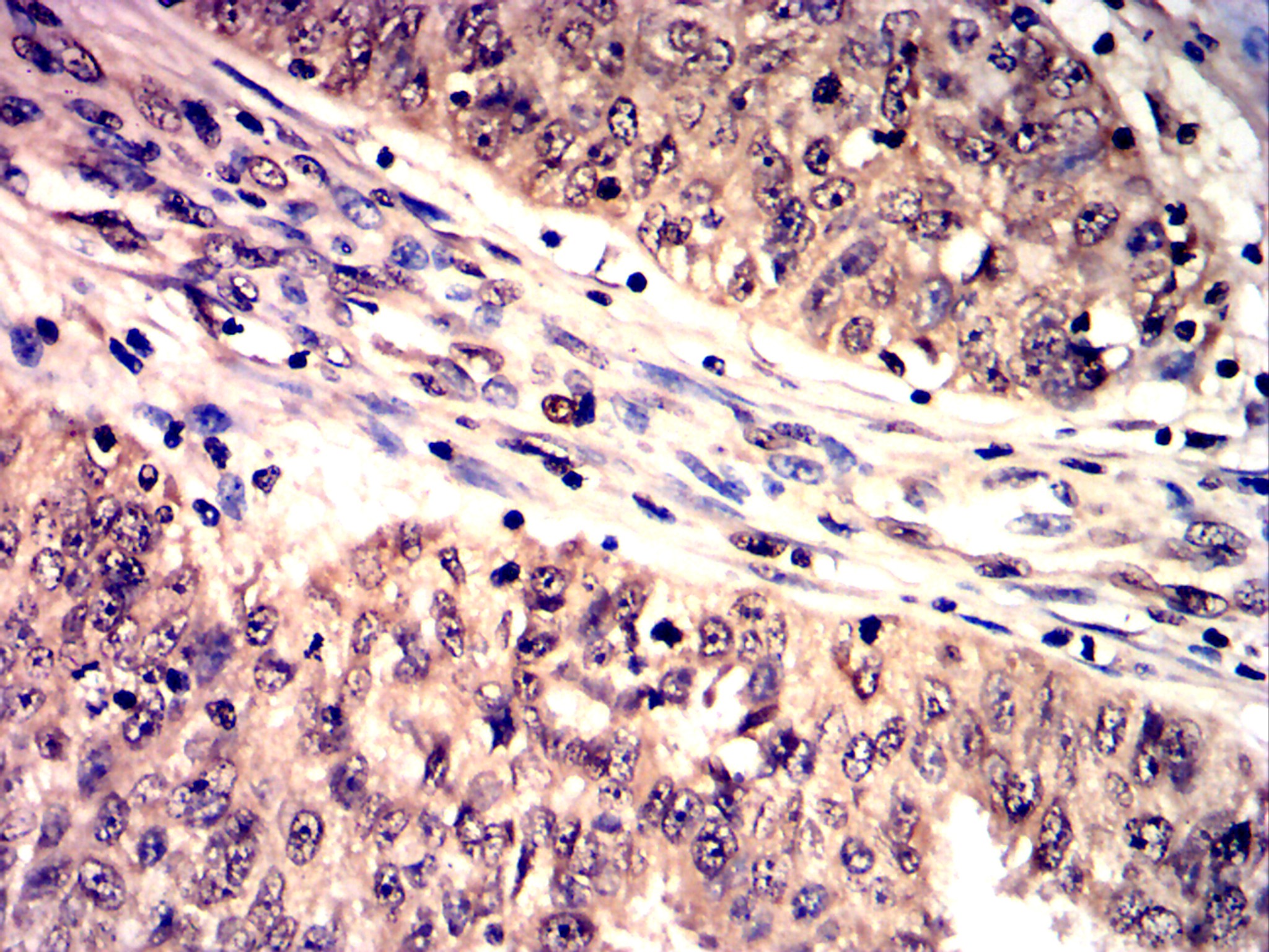
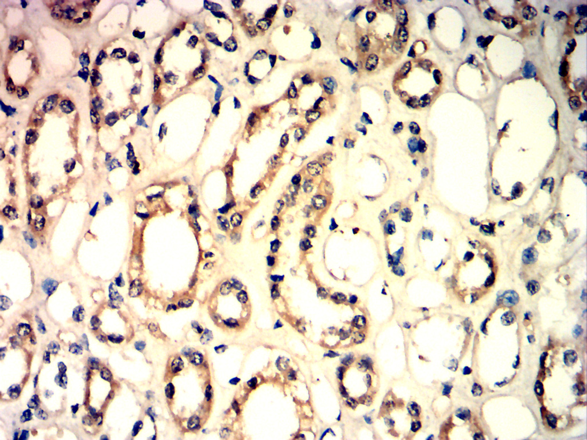
Immunohistochemical analysis of paraffin-embedded Human renal carcinoma tissues using Histone H3 antibody with DAB staining.IHC
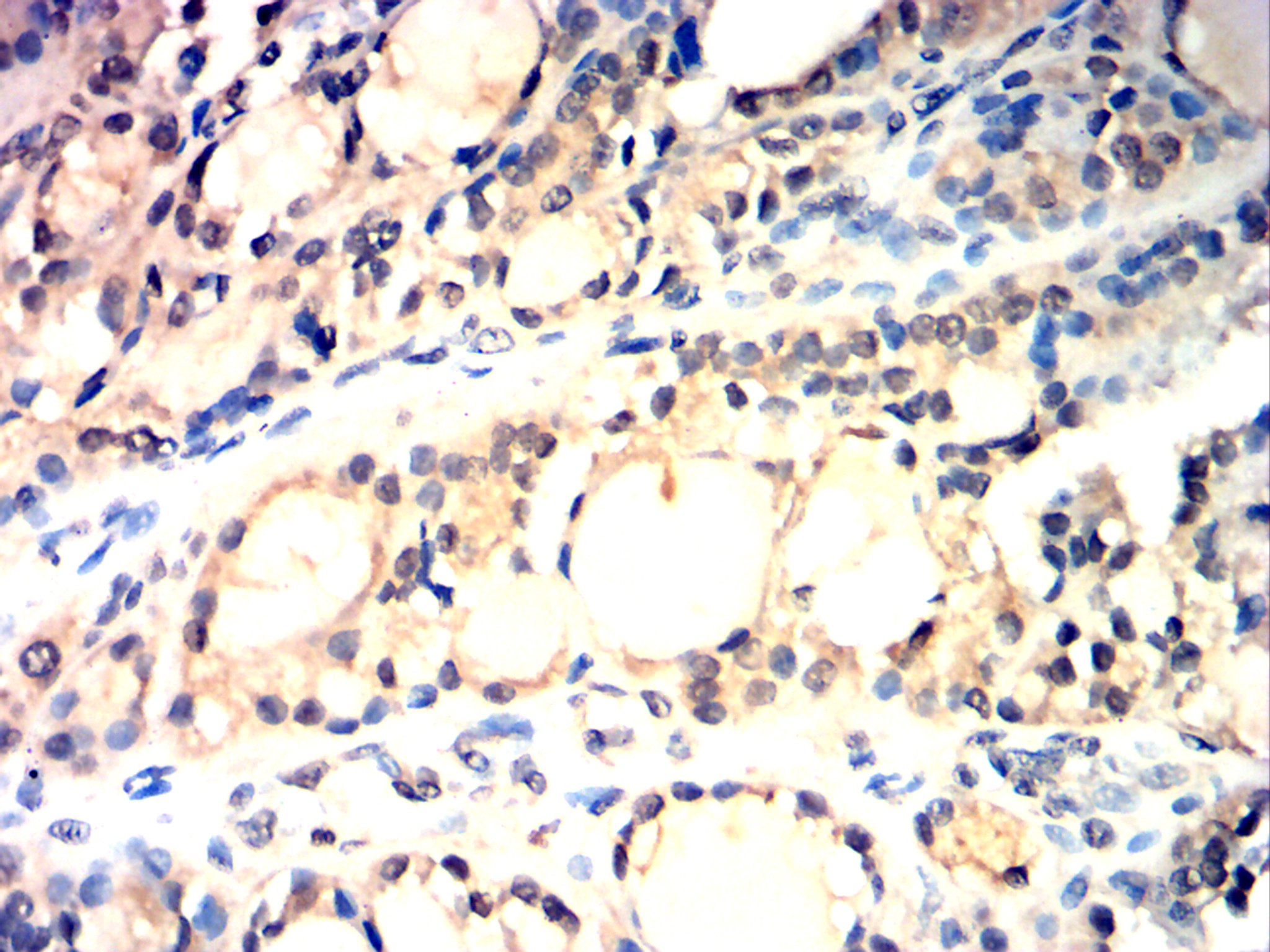
Immunohistochemical analysis of paraffin-embedded Human thyroid carcinoma tissues using Histone H3 antibody with DAB staining.| Product Name | Anti-Histone H3 antibody-HRP |
|---|---|
| Antibody Type | Primary Antibodies |
| Immunogen | Polypeptide |
| Conjugation | HRP |
| Clonality | Polyclonal |
|---|---|
| Isotype | IgG |
| Host Species | Rabbit |
| Tested Applications | WBIHC |
| WB:1:200-1:2000 IHC:1:100-1:1000 |
|
| Species Reactivity | HumanMouseRat |
| Concentration | 1mg/ml |
| Purification | Protein A |
| Gene Symbol | H3C1 |
|---|---|
| Gene Synonyms | H3 |
| Gene Full Name | H3 clustered histone 14 |
| Gene Summary | Histones are basic nuclear proteins that are responsible for the nucleosome structure of the chromosomal fiber in eukaryotes. This structure consists of approximately 146 bp of DNA wrapped around a nucleosome, an octamer composed of pairs of each of the four core histones (H2A, H2B, H3, and H4). The chromatin fiber is further compacted through the interaction of a linker histone, H1, with the DNA between the nucleosomes to form higher order chromatin structures. This gene is intronless and encodes a replication-dependent histone that is a member of the histone H3 family. Transcripts from this gene lack polyA tails; instead, they contain a palindromic termination element. This gene is found in a histone cluster on chromosome 1. This gene is one of four histone genes in the cluster that are duplicated; this record represents the telomeric copy. [provided by RefSeq, Aug 2015] |
| Molecular Weight(MW) | 15 kDa |
| Source | Rabbit |
| Cellular Localization | Nucleus |
WB
Western blot analysis using Histone H3 antibody against PANC-1 (1) cell lysate.15% SDS-PAGE gel.Sample loading: 20μg /lane. Transfer the proteins onto a PVDF membrane (OM790003), and block it with TBST (OM750016) plus skimmed milk powder for one hour. Dilute the primary antibody with the antibody diluent (OM750012) at a ratio of 1:1000, and incubate it overnight at 4°C. Wash the membrane three times with TBST (OM750016), 5 minutes each time. At room temperature, dilute the secondary antibody, Goat Anti-Rabbit IgG(H&L)-HRP (OM643487), at a ratio of 1:20000 and incubate for one hour. Wash the membrane three times with TBST (OM750016) again, 5 minutes each time. Use ECL (OM625701) for luminescence.
IHC
Immunohistochemical analysis of paraffin-embedded Human lung cancer tissues using Histone H3 antibody with DAB staining.
IHC
Immunohistochemical analysis of paraffin-embedded Human breast cancer tissues using Histone H3 antibody with DAB staining.
IHC
Immunohistochemical analysis of paraffin-embedded Human prostate cancer tissues using Histone H3 antibody with DAB staining.
IHC
Immunohistochemical analysis of paraffin-embedded Human ovarian cancer tissues using Histone H3 antibody with DAB staining.
IHC
Immunohistochemical analysis of paraffin-embedded Human renal carcinoma tissues using Histone H3 antibody with DAB staining.
IHC
Immunohistochemical analysis of paraffin-embedded Human thyroid carcinoma tissues using Histone H3 antibody with DAB staining.| Application Notes | WB:1:200-1:2000 IHC:1:100-1:1000 |
|---|
| Form | Liquid |
|---|---|
| Storage Instructions | Shipped at 4°C. Store at +4°C short term (1-2 weeks). Store at -20°C long term. Avoid freeze / thaw cycle. |
| Storage Buffer | Purified antibody in PBS with 0.05% sodium azide. |


