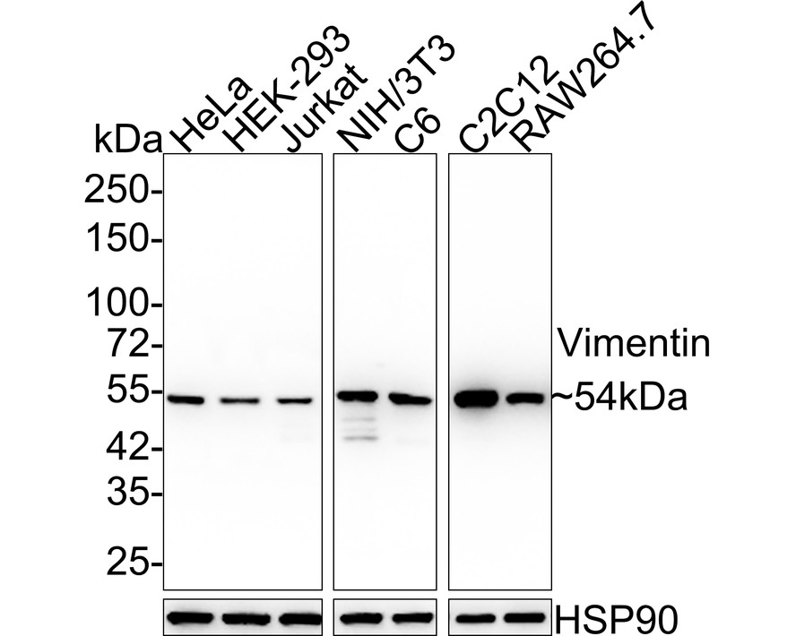
WB
Western blot analysis of Vimentin on different lysates with Rabbit anti-Vimentin antibody at 1/20,000 dilution. Lane 1: HeLa cell lysate (10 µg/Lane) Lane 2: HEK-293 cell lysate (10 µg/Lane) Lane 3: Jurkat cell lysate (10 µg/Lane) Lane 4: NIH/3T3 cell lysate (10 µg/Lane) Lane 5: C6 cell lysate (10 µg/Lane) Lane 6: C2C12 cell lysate (10 µg/Lane) Lane 7: RAW264.7 cell lysate (10 µg/Lane) Predicted band size: 54 kDa Observed band size: 54 kDa Exposure time: Lane 1-5: 3 seconds; Lane 6-7: 14 seconds; 4-20% SDS-PAGE gel. Proteins were transferred to a PVDF membrane and blocked with 5% NFDM/TBST for 1 hour at room temperature. The primary antibody at 1/20,000 dilution was used in 5% NFDM/TBST at room temperature for 2 hours. Goat Anti-Rabbit IgG - HRP Secondary Antibody at 1:50,000 dilution was used for 1 hour at room temperature.IHC
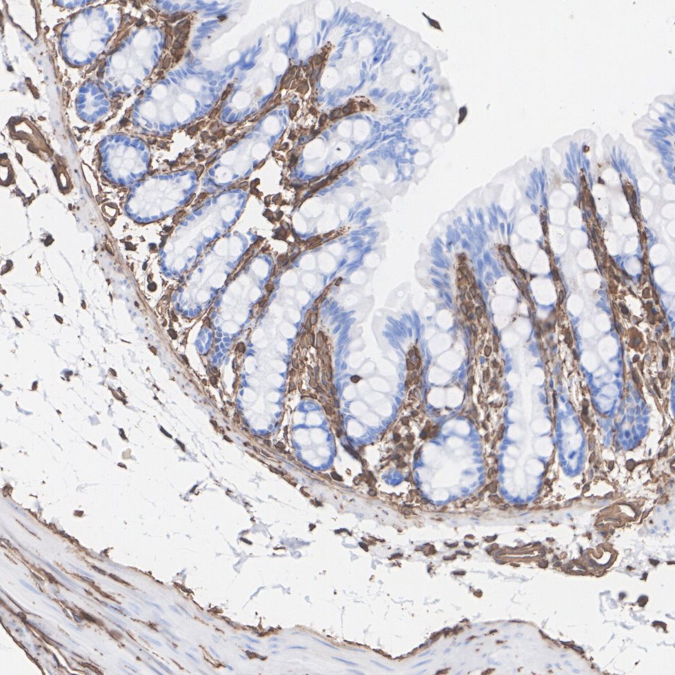
Immunohistochemical analysis of paraffin-embedded rat colon tissue with Rabbit anti-Vimentin antibody at 1/5,000 dilution. The section was pre-treated using heat mediated antigen retrieval with Tris-EDTA buffer (pH 9.0) for 20 minutes. The tissues were blocked in 1% BSA for 20 minutes at room temperature, washed with ddH2O and PBS, and then probed with the primary antibody at 1/5,000 dilution for 1 hour at room temperature. The detection was performed using an HRP conjugated compact polymer system. DAB was used as the chromogen. Tissues were counterstained with hematoxylin and mounted with DPX.ICC/IF
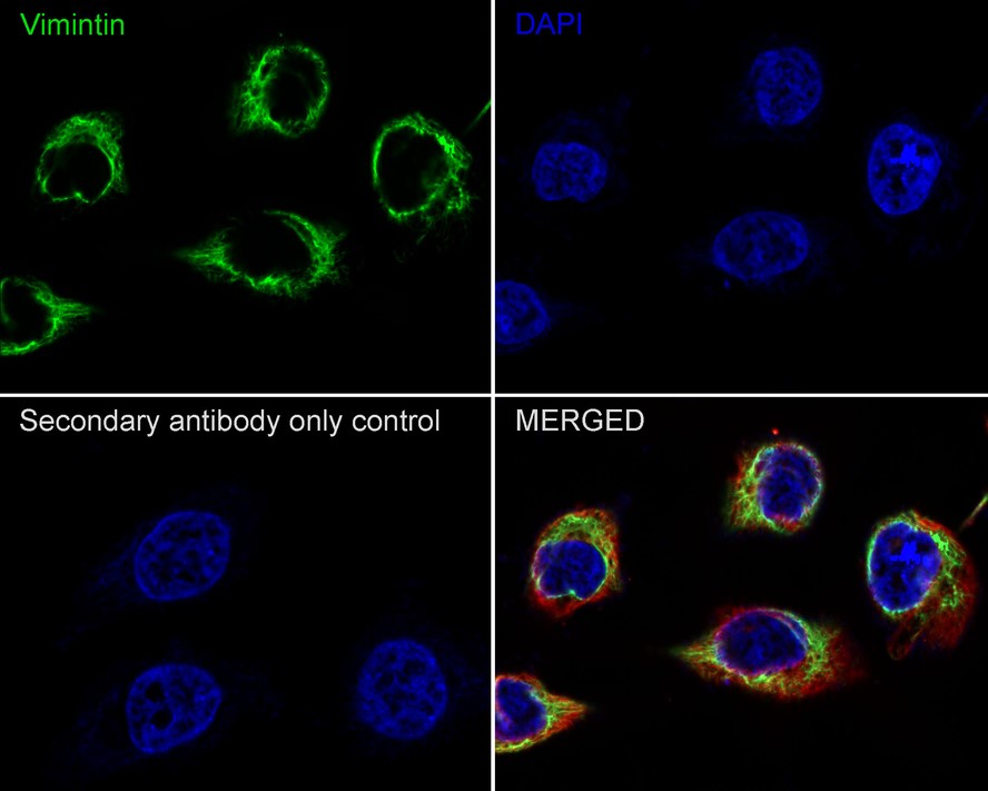
Immunocytochemistry analysis of HeLa cells labeling Vimentin with Rabbit anti-Vimentin antibody at 1/200 dilution. Cells were fixed in 4% paraformaldehyde for 10 minutes at 37 ℃, permeabilized with 0.05% Triton X-100 in PBS for 20 minutes, and then blocked with 2% negative goat serum for 30 minutes at room temperature. Cells were then incubated with Rabbit anti-Vimentin antibody at 1/200 dilution in 2% negative goat serum overnight at 4 ℃. Goat Anti-Rabbit IgG H&L (iFluor™ 488) was used as the secondary antibody at 1/1,000 dilution. Nuclear DNA was labelled in blue with DAPI. Beta tubulin ( red) was stained at 1/100 dilution overnight at +4℃. Goat Anti-Mouse IgG H&L (iFluor™ 594) was used as the secondary antibody at 1/1,000 dilution.FC
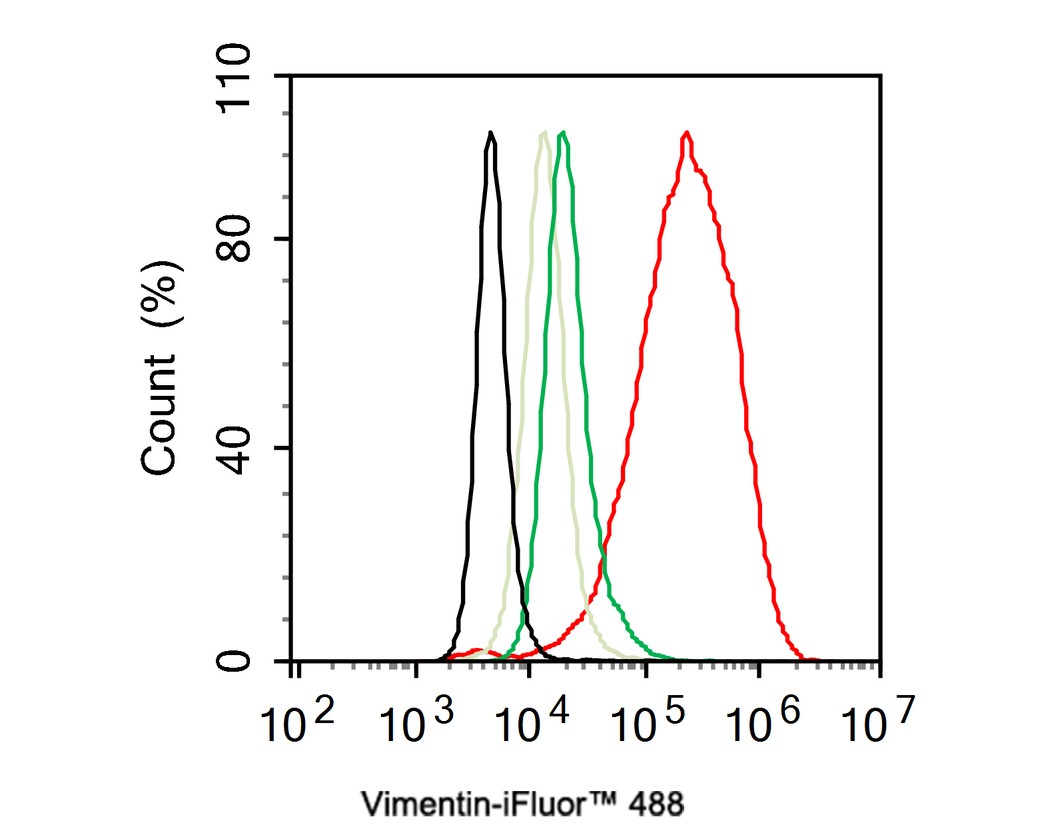
Flow cytometric analysis of HeLa (positive, red) and MCF7 (negative, green) cells labeling Vimentin. Cells were fixed by 4% formaldehyde and then permeabilized with ice-cold 90% methanol. Then stained with the primary antibody at 1/1,000 dilution, compared with Rabbit IgG Isotype Control (HeLa black, MCF7 light green). After incubation of the primary antibody at +4℃ for an hour, the cells were stained with a iFluor™ 488 conjugate-Goat anti-Rabbit IgG Secondary antibody at 1/1,000 dilution for 30 minutes at +4℃.IP
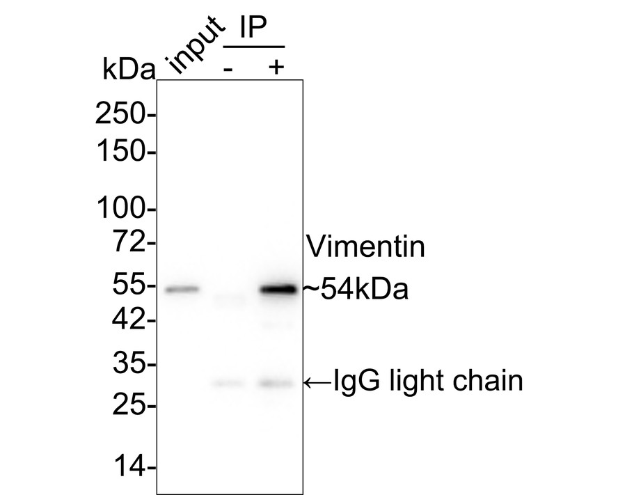
Vimentin was immunoprecipitated in 0.2mg HeLa cell lysate with Rabbit anti-Vimentin antibody at 2 µg/25 µl agarose. Western blot was performed from the immunoprecipitate using Rabbit anti-Vimentin antibody at 1/10,000 dilution. Anti-Rabbit IgG for IP Nano-secondary antibody at 1/5,000 dilution was used for 1 hour at room temperature. Lane 1: HeLa cell lysate (input), Lane 2: Rabbit IgG instead of Rabbit anti-Vimentin antibody in HeLa cell lysate, Lane 3: Rabbit anti-Vimentin antibody IP in HeLa cell lysate. Blocking/Dilution buffer: 5% NFDM/TBST. Exposure time: 5 seconds.IF-P
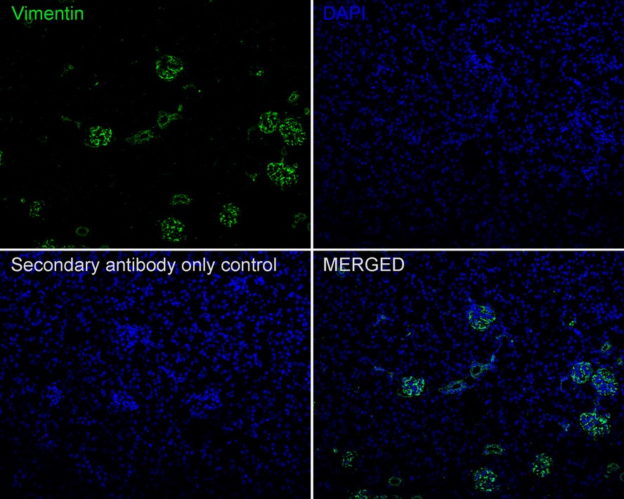
Immunofluorescence analysis of paraffin-embedded mouse kidney tissue labeling Vimentin with Rabbit anti-Vimentin antibody at 1/1,000 dilution. The section was pre-treated using heat mediated antigen retrieval with Tris-EDTA buffer (pH 9.0) for 20 minutes. The tissues were blocked in 10% negative goat serum for 1 hour at room temperature, washed with PBS, and then probed with the primary antibody (green) at 1/1,000 dilution overnight at 4 ℃, washed with PBS. Goat Anti-Rabbit IgG H&L (iFluor™ 488) was used as the secondary antibody at 1/1,000 dilution. Nuclei were counterstained with DAPI (blue).| Product Name | Vimentin Recombinant Rabbit Monoclonal Antibody |
|---|---|
| Antibody Type | Primary Antibodies |
| Immunogen | Synthetic peptide within C-terminal human Vimentin. |
| Isotype | IgG |
|---|---|
| Host Species | Rabbit |
| Tested Applications | FCICC/IFIF-PIHCIPWB |
| WB:120000 IHC:1:100-1:10000 ICC:1:100-1:500 FC:1:50-1:100 |
|
| Species Reactivity | HumanMouseRat |
| Concentration | 1mg/ml |
| Purification | Protein A |
| Gene Symbol | VIM |
|---|---|
| Gene Full Name | vimentin |
| Gene Summary | This gene encodes a type III intermediate filament protein. Intermediate filaments, along with microtubules and actin microfilaments, make up the cytoskeleton. The encoded protein is responsible for maintaining cell shape and integrity of the cytoplasm, and stabilizing cytoskeletal interactions. This protein is involved in neuritogenesis and cholesterol transport and functions as an organizer of a number of other critical proteins involved in cell attachment, migration, and signaling. Bacterial and viral pathogens have been shown to attach to this protein on the host cell surface. Mutations in this gene are associated with congenital cataracts in human patients. [provided by RefSeq, Aug 2017] |
| Molecular Weight(MW) | 54kDa |
| Cellular Localization | Cytoplasm. |
WB
Western blot analysis of Vimentin on different lysates with Rabbit anti-Vimentin antibody at 1/20,000 dilution. Lane 1: HeLa cell lysate (10 µg/Lane) Lane 2: HEK-293 cell lysate (10 µg/Lane) Lane 3: Jurkat cell lysate (10 µg/Lane) Lane 4: NIH/3T3 cell lysate (10 µg/Lane) Lane 5: C6 cell lysate (10 µg/Lane) Lane 6: C2C12 cell lysate (10 µg/Lane) Lane 7: RAW264.7 cell lysate (10 µg/Lane) Predicted band size: 54 kDa Observed band size: 54 kDa Exposure time: Lane 1-5: 3 seconds; Lane 6-7: 14 seconds; 4-20% SDS-PAGE gel. Proteins were transferred to a PVDF membrane and blocked with 5% NFDM/TBST for 1 hour at room temperature. The primary antibody at 1/20,000 dilution was used in 5% NFDM/TBST at room temperature for 2 hours. Goat Anti-Rabbit IgG - HRP Secondary Antibody at 1:50,000 dilution was used for 1 hour at room temperature.
IHC
Immunohistochemical analysis of paraffin-embedded rat colon tissue with Rabbit anti-Vimentin antibody at 1/5,000 dilution. The section was pre-treated using heat mediated antigen retrieval with Tris-EDTA buffer (pH 9.0) for 20 minutes. The tissues were blocked in 1% BSA for 20 minutes at room temperature, washed with ddH2O and PBS, and then probed with the primary antibody at 1/5,000 dilution for 1 hour at room temperature. The detection was performed using an HRP conjugated compact polymer system. DAB was used as the chromogen. Tissues were counterstained with hematoxylin and mounted with DPX.
ICC/IF
Immunocytochemistry analysis of HeLa cells labeling Vimentin with Rabbit anti-Vimentin antibody at 1/200 dilution. Cells were fixed in 4% paraformaldehyde for 10 minutes at 37 ℃, permeabilized with 0.05% Triton X-100 in PBS for 20 minutes, and then blocked with 2% negative goat serum for 30 minutes at room temperature. Cells were then incubated with Rabbit anti-Vimentin antibody at 1/200 dilution in 2% negative goat serum overnight at 4 ℃. Goat Anti-Rabbit IgG H&L (iFluor™ 488) was used as the secondary antibody at 1/1,000 dilution. Nuclear DNA was labelled in blue with DAPI. Beta tubulin ( red) was stained at 1/100 dilution overnight at +4℃. Goat Anti-Mouse IgG H&L (iFluor™ 594) was used as the secondary antibody at 1/1,000 dilution.
FC
Flow cytometric analysis of HeLa (positive, red) and MCF7 (negative, green) cells labeling Vimentin. Cells were fixed by 4% formaldehyde and then permeabilized with ice-cold 90% methanol. Then stained with the primary antibody at 1/1,000 dilution, compared with Rabbit IgG Isotype Control (HeLa black, MCF7 light green). After incubation of the primary antibody at +4℃ for an hour, the cells were stained with a iFluor™ 488 conjugate-Goat anti-Rabbit IgG Secondary antibody at 1/1,000 dilution for 30 minutes at +4℃.
IP
Vimentin was immunoprecipitated in 0.2mg HeLa cell lysate with Rabbit anti-Vimentin antibody at 2 µg/25 µl agarose. Western blot was performed from the immunoprecipitate using Rabbit anti-Vimentin antibody at 1/10,000 dilution. Anti-Rabbit IgG for IP Nano-secondary antibody at 1/5,000 dilution was used for 1 hour at room temperature. Lane 1: HeLa cell lysate (input), Lane 2: Rabbit IgG instead of Rabbit anti-Vimentin antibody in HeLa cell lysate, Lane 3: Rabbit anti-Vimentin antibody IP in HeLa cell lysate. Blocking/Dilution buffer: 5% NFDM/TBST. Exposure time: 5 seconds.
IF-P
Immunofluorescence analysis of paraffin-embedded mouse kidney tissue labeling Vimentin with Rabbit anti-Vimentin antibody at 1/1,000 dilution. The section was pre-treated using heat mediated antigen retrieval with Tris-EDTA buffer (pH 9.0) for 20 minutes. The tissues were blocked in 10% negative goat serum for 1 hour at room temperature, washed with PBS, and then probed with the primary antibody (green) at 1/1,000 dilution overnight at 4 ℃, washed with PBS. Goat Anti-Rabbit IgG H&L (iFluor™ 488) was used as the secondary antibody at 1/1,000 dilution. Nuclei were counterstained with DAPI (blue).| Application Notes | WB:120000 IHC:1:100-1:10000 ICC:1:100-1:500 FC:1:50-1:100 |
|---|
| Form | Liquid |
|---|---|
| Storage Instructions | Store at +4℃ after thawing. Aliquot store at -20℃ or -80℃. Avoid repeated freeze / thaw cycles. |
| Storage Buffer | 1*TBS (pH7.4), 0.05% BSA, 40% Glycerol. Preservative: 0.05% Sodium Azide. |


 Data sheet for OM642894