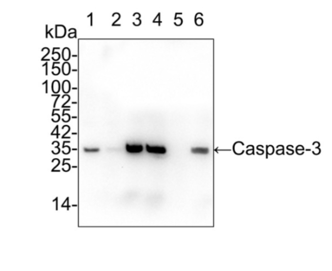
WB
Western blot analysis of Caspase-3 on different lysates with Rabbit anti-Caspase-3 antibody at 1/2,000 dilution. Lane 1: HeLa cell lysate Lane 2: HeLa treated with 1μM staurosporine for 3 hours cell lysate Lane 3: Jurkat cell lysate Lane 4: Jurkat treated with 25μM Etoposide for 5 hours cell lysate Lane 5: MCF7 cell lysate (negative) Lane 6: HEK-293 cell lysate Lysates/proteins at 20 µg/Lane. Predicted band size: 32 kDa Observed band size: 32 kDa Exposure time: 3 minutes 20 seconds; 4-20% SDS-PAGE gel. Proteins were transferred to a PVDF membrane and blocked with 5% NFDM/TBST for 1 hour at room temperature. The primary antibody at 1/2,000 dilution was used in 5% NFDM/TBST at 4℃ overnight. Goat Anti-Rabbit IgG - HRP Secondary Antibody at 1/50,000 dilution was used for 1 hour at room temperature.IHC
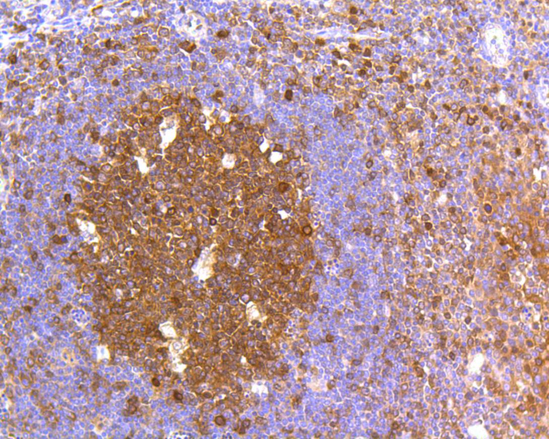
Immunohistochemical analysis of paraffin-embedded human tonsil tissue using anti-Caspase-3 antibody. The section was pre-treated using heat mediated antigen retrieval with Tris-EDTA buffer (pH 8.0-8.4) for 20 minutes.The tissues were blocked in 5% BSA for 30 minutes at room temperature, washed with ddH2O and PBS, and then probed with the primary antibody (1/50) for 30 minutes at room temperature. The detection was performed using an HRP conjugated compact polymer system. DAB was used as the chromogen. Tissues were counterstained with hematoxylin and mounted with DPX.ICC/IF
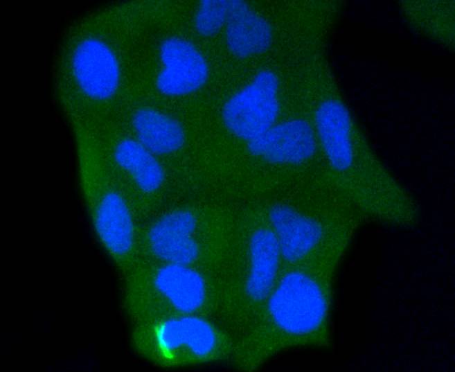
ICC staining of Caspase-3 in Hela cells (green). Formalin fixed cells were permeabilized with 0.1% Triton X-100 in TBS for 10 minutes at room temperature and blocked with 1% Blocker BSA for 15 minutes at room temperature. Cells were probed with the primary antibody (1/50) for 1 hour at room temperature, washed with PBS. Alexa Fluor®488 Goat anti-Rabbit IgG was used as the secondary antibody at 1/1,000 dilution. The nuclear counter stain is DAPI (blue).| Product Name | Caspase-3 Recombinant Rabbit Monoclonal Antibody |
|---|---|
| Antibody Type | Primary Antibodies |
| Immunogen | Synthetic peptide within human Caspase-3 aa 60-100. |
| Clonality | monoclonal |
|---|---|
| Isotype | IgG |
| Host Species | Rabbit |
| Tested Applications | FCICC/IFIHCIPWB |
| WB:1:2000-1:5000 IHC:1:50 ICC:1:50 FC:1:20-1:50 IP:Use at an assay dependent concentration. |
|
| Species Reactivity | Human |
| Concentration | 1mg/ml |
| Purification | Protein A |
| Gene Symbol | CASP3 |
|---|---|
| Gene Synonyms | CPP32 SCA-1 CPP32B |
| Gene Full Name | caspase 3 |
| Gene Summary | The protein encoded by this gene is a cysteine-aspartic acid protease that plays a central role in the execution-phase of cell apoptosis. The encoded protein cleaves and inactivates poly(ADP-ribose) polymerase while it cleaves and activates sterol regulatory element binding proteins as well as caspases 6, 7, and 9. This protein itself is processed by caspases 8, 9, and 10. It is the predominant caspase involved in the cleavage of amyloid-beta 4A precursor protein, which is associated with neuronal death in Alzheimer's disease. [provided by RefSeq, Aug 2017] |
| Molecular Weight(MW) | 32kDa |
| Cellular Localization | Cytoplasm. |
WB
Western blot analysis of Caspase-3 on different lysates with Rabbit anti-Caspase-3 antibody at 1/2,000 dilution. Lane 1: HeLa cell lysate Lane 2: HeLa treated with 1μM staurosporine for 3 hours cell lysate Lane 3: Jurkat cell lysate Lane 4: Jurkat treated with 25μM Etoposide for 5 hours cell lysate Lane 5: MCF7 cell lysate (negative) Lane 6: HEK-293 cell lysate Lysates/proteins at 20 µg/Lane. Predicted band size: 32 kDa Observed band size: 32 kDa Exposure time: 3 minutes 20 seconds; 4-20% SDS-PAGE gel. Proteins were transferred to a PVDF membrane and blocked with 5% NFDM/TBST for 1 hour at room temperature. The primary antibody at 1/2,000 dilution was used in 5% NFDM/TBST at 4℃ overnight. Goat Anti-Rabbit IgG - HRP Secondary Antibody at 1/50,000 dilution was used for 1 hour at room temperature.
IHC
Immunohistochemical analysis of paraffin-embedded human tonsil tissue using anti-Caspase-3 antibody. The section was pre-treated using heat mediated antigen retrieval with Tris-EDTA buffer (pH 8.0-8.4) for 20 minutes.The tissues were blocked in 5% BSA for 30 minutes at room temperature, washed with ddH2O and PBS, and then probed with the primary antibody (1/50) for 30 minutes at room temperature. The detection was performed using an HRP conjugated compact polymer system. DAB was used as the chromogen. Tissues were counterstained with hematoxylin and mounted with DPX.
ICC/IF
ICC staining of Caspase-3 in Hela cells (green). Formalin fixed cells were permeabilized with 0.1% Triton X-100 in TBS for 10 minutes at room temperature and blocked with 1% Blocker BSA for 15 minutes at room temperature. Cells were probed with the primary antibody (1/50) for 1 hour at room temperature, washed with PBS. Alexa Fluor®488 Goat anti-Rabbit IgG was used as the secondary antibody at 1/1,000 dilution. The nuclear counter stain is DAPI (blue).| Application Notes | WB:1:2000-1:5000 IHC:1:50 ICC:1:50 FC:1:20-1:50 IP:Use at an assay dependent concentration. |
|---|
| Form | Liquid |
|---|---|
| Storage Instructions | Store at +4℃ after thawing. Aliquot store at -20℃ or -80℃. Avoid repeated freeze / thaw cycles. |
| Storage Buffer | 1*TBS (pH7.4), 0.05% BSA, 40% Glycerol. Preservative: 0.05% Sodium Azide. |


 Data sheet for OM643026