WB
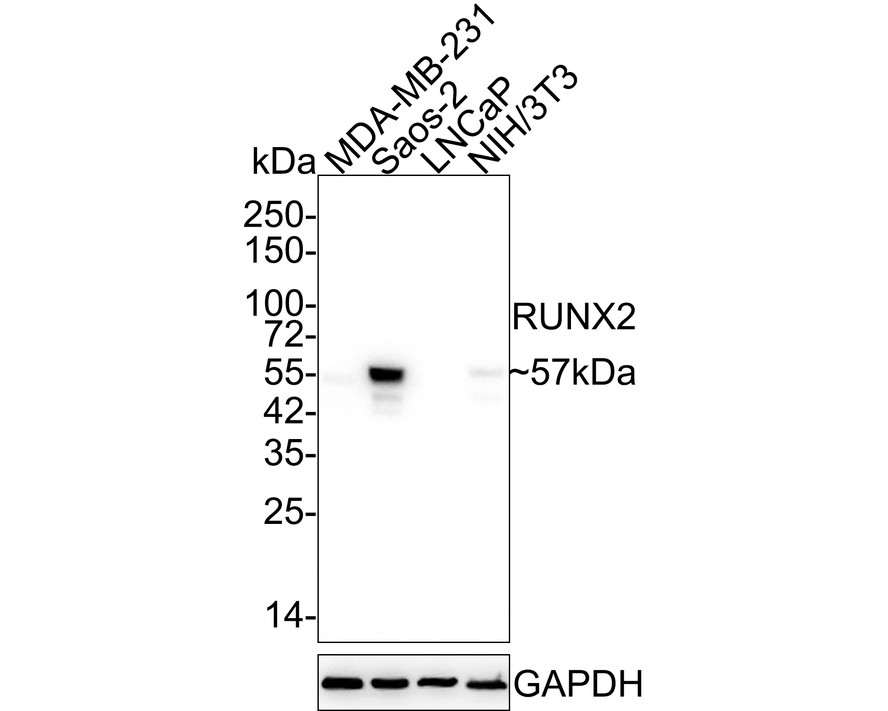
Western blot analysis of RUNX2 on different lysates with Rabbit anti-RUNX2 antibody at 1/10,000 dilution. Lane 1: MDA-MB-231 cell lysate, Lane 2: Saos-2 cell lysate, Lane 3: LNCaP cell lysate (low expression), Lane 4: NIH/3T3 cell lysate, Lysates/proteins at 20 µg/Lane. Exposure time: 2 minutes 24 seconds; 4-20% SDS-PAGE gel. Proteins were transferred to a PVDF membrane and blocked with 5% NFDM/TBST for 1 hour at room temperature. The primary antibody at 1/10,000 dilution was used in 5% NFDM/TBST at 4℃ overnight. Goat Anti-Rabbit IgG - HRP Secondary Antibody at 1/50,000 dilution was used for 1 hour at room temperature.IHC
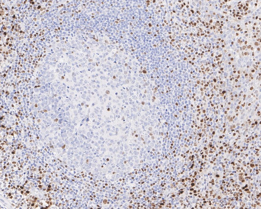
Immunohistochemical analysis of paraffin-embedded human tonsil tissue with Rabbit anti-RUNX2 antibody at 1/1,000 dilution. The section was pre-treated using heat mediated antigen retrieval with sodium citrate buffer (pH 6.0) for 2 minutes. The tissues were blocked in 1% BSA for 20 minutes at room temperature, washed with ddH2O and PBS, and then probed with the primary antibody at 1/1,000 dilution for 1 hour at room temperature. The detection was performed using an HRP conjugated compact polymer system. DAB was used as the chromogen. Tissues were counterstained with hematoxylin and mounted with DPX.ICC/IF
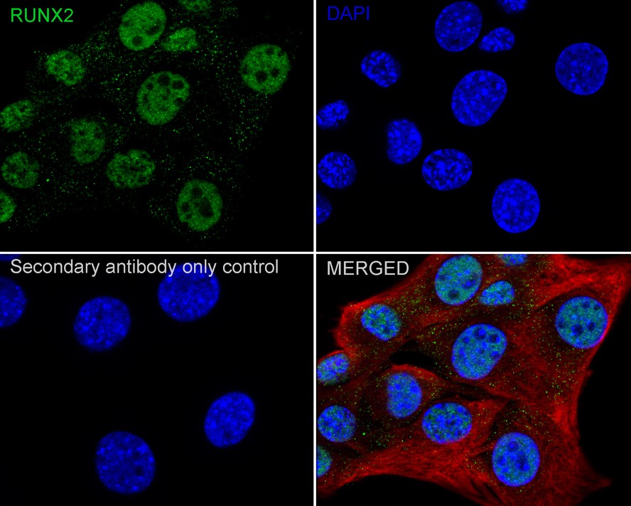
Immunocytochemistry analysis of C2C12 cells labeling RUNX2 with Rabbit anti-RUNX2 antibody at 1/1,000 dilution. Cells were fixed in 4% paraformaldehyde for 15 minutes at room temperature, permeabilized with 0.1% Triton X-100 in PBS for 15 minutes at room temperature, then blocked with 1% BSA in 10% negative goat serum for 1 hour at room temperature. Cells were then incubated with Rabbit anti-RUNX2 antibody at 1/1,000 dilution in 1% BSA in PBST overnight at 4 ℃. Goat Anti-Rabbit IgG H&L (iFluor™ 488) was used as the secondary antibody at 1/1,000 dilution. PBS instead of the primary antibody was used as the secondary antibody only control. Nuclear DNA was labelled in blue with DAPI. Beta tubulin (red) was stained at 1/100 dilution overnight at +4℃. Goat Anti-Mouse IgG H&L (iFluor™ 594) was used as the secondary antibody at 1/1,000 dilution.FC
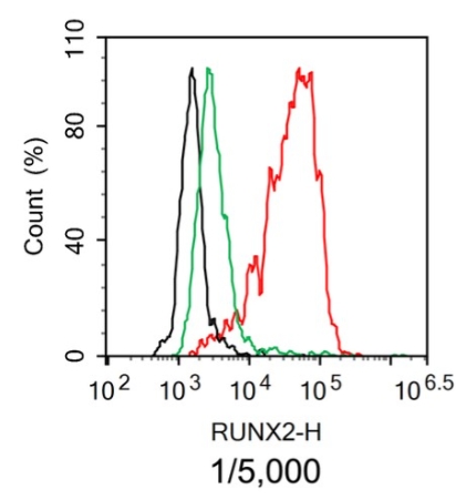
Flow cytometric analysis of Saos-2 cells labeling RUNX2. Cells were fixed and permeabilized. Then stained with the primary antibody (red) at 1/5,000 dilution , compared with Rabbit IgG Isotype Control (green). After incubation of the primary antibody at +4℃ for an hour, the cells were stained with a iFluor™ 488 conjugate-Goat anti-Rabbit IgG Secondary antibody at 1/1,000 dilution for 30 minutes at +4℃. Unlabelled sample was used as a control (cells without incubation with primary antibody; black).IP
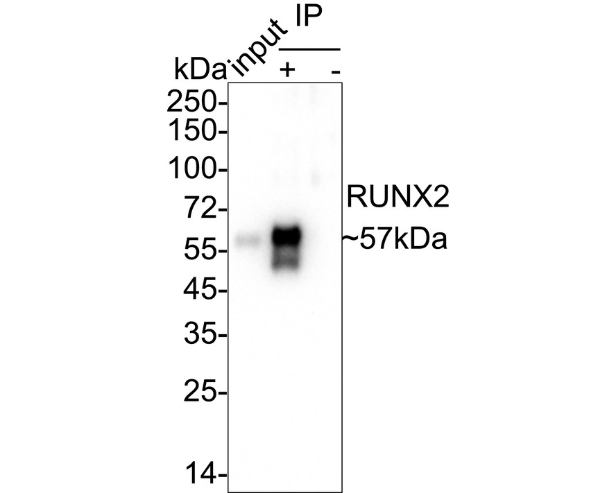
RUNX2 was immunoprecipitated from 0.2 mg Saos-2 cell lysate with Rabbit anti-RUNX2 antibody at 2 µg/25 µl agarose. Western blot was performed from the immunoprecipitate using Rabbit anti-RUNX2 antibody at 1/10,000 dilution. Anti-Rabbit IgG for IP Nano-secondary antibody at 1/5,000 dilution was used for 1 hour at room temperature. Lane 1: Saos-2 cell lysate (input) Lane 2: Rabbit anti-RUNX2 antibody IP in Saos-2 cell lysate Lane 3: Rabbit IgG instead of Rabbit anti-RUNX2 antibody in Saos-2 cell lysate Blocking/Dilution buffer: 5% NFDM/TBST Exposure time: 20 seconds.| Product Name | RUNX2 Recombinant Rabbit Monoclonal Antibody |
|---|---|
| Antibody Type | Primary Antibodies |
| Immunogen | Recombinant protein within human 300-450. |
| Clonality | Monoclonal |
|---|---|
| Isotype | IgG |
| Host Species | Rabbit |
| Tested Applications | FCICC/IFIHCIPWB |
| WB:1:5000-1:10000 IHC:1:200-1:1000 ICC:1:1000-1:5000 FC:1:5000 IP:1-2μg/sample |
|
| Species Reactivity | HumanMouseRat |
| Concentration | 1mg/ml |
| Purification | Protein A |
| Gene Symbol | RUNX2 |
|---|---|
| Gene Synonyms | CCD AML3 CCD1 CLCD OSF2 CBFA1 OSF-2 PEA2aA PEBP2aA CBF-alpha-1 |
| Gene Full Name | RUNX family transcription factor 2 |
| Gene Summary | This gene is a member of the RUNX family of transcription factors and encodes a nuclear protein with an Runt DNA-binding domain. This protein is essential for osteoblastic differentiation and skeletal morphogenesis and acts as a scaffold for nucleic acids and regulatory factors involved in skeletal gene expression. The protein can bind DNA both as a monomer or, with more affinity, as a subunit of a heterodimeric complex. Two regions of potential trinucleotide repeat expansions are present in the N-terminal region of the encoded protein, and these and other mutations in this gene have been associated with the bone development disorder cleidocranial dysplasia (CCD). Transcript variants that encode different protein isoforms result from the use of alternate promoters as well as alternate splicing. [provided by RefSeq, Jul 2016] |
| Molecular Weight(MW) | 57kDa |
| Cellular Localization | Nucleus. |
WB
Western blot analysis of RUNX2 on different lysates with Rabbit anti-RUNX2 antibody at 1/10,000 dilution. Lane 1: MDA-MB-231 cell lysate, Lane 2: Saos-2 cell lysate, Lane 3: LNCaP cell lysate (low expression), Lane 4: NIH/3T3 cell lysate, Lysates/proteins at 20 µg/Lane. Exposure time: 2 minutes 24 seconds; 4-20% SDS-PAGE gel. Proteins were transferred to a PVDF membrane and blocked with 5% NFDM/TBST for 1 hour at room temperature. The primary antibody at 1/10,000 dilution was used in 5% NFDM/TBST at 4℃ overnight. Goat Anti-Rabbit IgG - HRP Secondary Antibody at 1/50,000 dilution was used for 1 hour at room temperature.
IHC
Immunohistochemical analysis of paraffin-embedded human tonsil tissue with Rabbit anti-RUNX2 antibody at 1/1,000 dilution. The section was pre-treated using heat mediated antigen retrieval with sodium citrate buffer (pH 6.0) for 2 minutes. The tissues were blocked in 1% BSA for 20 minutes at room temperature, washed with ddH2O and PBS, and then probed with the primary antibody at 1/1,000 dilution for 1 hour at room temperature. The detection was performed using an HRP conjugated compact polymer system. DAB was used as the chromogen. Tissues were counterstained with hematoxylin and mounted with DPX.
ICC/IF
Immunocytochemistry analysis of C2C12 cells labeling RUNX2 with Rabbit anti-RUNX2 antibody at 1/1,000 dilution. Cells were fixed in 4% paraformaldehyde for 15 minutes at room temperature, permeabilized with 0.1% Triton X-100 in PBS for 15 minutes at room temperature, then blocked with 1% BSA in 10% negative goat serum for 1 hour at room temperature. Cells were then incubated with Rabbit anti-RUNX2 antibody at 1/1,000 dilution in 1% BSA in PBST overnight at 4 ℃. Goat Anti-Rabbit IgG H&L (iFluor™ 488) was used as the secondary antibody at 1/1,000 dilution. PBS instead of the primary antibody was used as the secondary antibody only control. Nuclear DNA was labelled in blue with DAPI. Beta tubulin (red) was stained at 1/100 dilution overnight at +4℃. Goat Anti-Mouse IgG H&L (iFluor™ 594) was used as the secondary antibody at 1/1,000 dilution.
FC
Flow cytometric analysis of Saos-2 cells labeling RUNX2. Cells were fixed and permeabilized. Then stained with the primary antibody (red) at 1/5,000 dilution , compared with Rabbit IgG Isotype Control (green). After incubation of the primary antibody at +4℃ for an hour, the cells were stained with a iFluor™ 488 conjugate-Goat anti-Rabbit IgG Secondary antibody at 1/1,000 dilution for 30 minutes at +4℃. Unlabelled sample was used as a control (cells without incubation with primary antibody; black).
IP
RUNX2 was immunoprecipitated from 0.2 mg Saos-2 cell lysate with Rabbit anti-RUNX2 antibody at 2 µg/25 µl agarose. Western blot was performed from the immunoprecipitate using Rabbit anti-RUNX2 antibody at 1/10,000 dilution. Anti-Rabbit IgG for IP Nano-secondary antibody at 1/5,000 dilution was used for 1 hour at room temperature. Lane 1: Saos-2 cell lysate (input) Lane 2: Rabbit anti-RUNX2 antibody IP in Saos-2 cell lysate Lane 3: Rabbit IgG instead of Rabbit anti-RUNX2 antibody in Saos-2 cell lysate Blocking/Dilution buffer: 5% NFDM/TBST Exposure time: 20 seconds.| Application Notes | WB:1:5000-1:10000 IHC:1:200-1:1000 ICC:1:1000-1:5000 FC:1:5000 IP:1-2μg/sample |
|---|
| Form | Liquid |
|---|---|
| Storage Instructions | Store at +4℃ after thawing. Aliquot store at -20℃ or -80℃. Avoid repeated freeze / thaw cycles. |
| Storage Buffer | 1*TBS (pH7.4), 0.05% BSA, 40% Glycerol. Preservative: 0.05% Sodium Azide. |


 Data sheet for OM643303