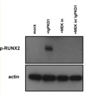
WB
Effect of IgPKD1, MEK inhibitor and dual IgPKD1/MEK inhibitor treatment on phosphorylated RUNX2 (p‐RUNX2) in trigonocephaly cranial suture cells.IHC
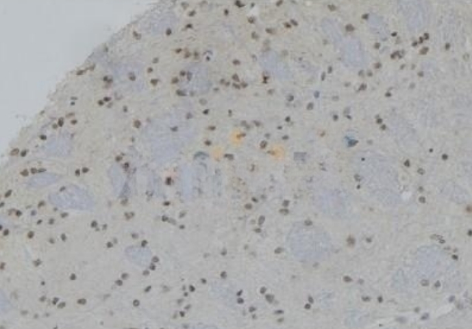
Phospho-RUNX2 (Ser275) Antibody at 1/100 staining rat brain tissue sections by IHC-P. The tissue was formaldehyde fixed and a heat mediated antigen retrieval step in citrate buffer was performed. The tissue was then blocked and incubated with the antibody for 1.5 hours at 22°C. An HRP conjugated goat anti-rabbit antibody was used as the secondary antibody.ICC/IF
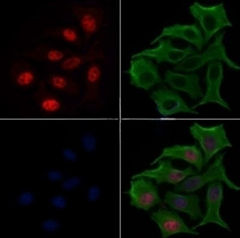
Phospho-RUNX2 (Ser275) Antibody staining HepG2 cells(serum starvation) by IF/ICC. The samples were fixed with PFA and permeabilized in 0.1% Triton X-100,then blocked in 10% serum for 45 minutes at 25°C. Samples were then incubated with primary Ab(1:200) and mouse anti-beta tubulin Ab(1:200) for 1 hour at 37°C. An AlexaFluor594 conjugated goat anti-rabbit IgG(H+L) Ab(Red) and an AlexaFluor488 conjugated goat anti-mouse IgG(H+L) Ab(Green) were used as the secondary antibody. The nuclear counter stain is DAPI(blue).| Product Name | Rabbit polyclonal antibody to Phospho-RUNX2 (Ser275) |
|---|---|
| Antibody Type | Primary Antibodies |
| Immunogen | A synthesized peptide derived from human RUNX2(Accession Q13950), corresponding to amino acid residues around phosphorylated Ser275. |
| Modification | p-Ser275 |
| Clonality | Polyclonal |
|---|---|
| Isotype | IgG |
| Host Species | Rabbit |
| Tested Applications | ICC/IFIHCWB |
| WB:1:500-1:2000 IHC: 1:50-1:200 ICC:1:100-1:500 |
|
| Species Reactivity | HumanMouseRat |
| Concentration | 1mg/ml |
| Purification | Affinity purified |
| Gene Symbol | RUNX2 |
|---|---|
| Gene Synonyms | CCD AML3 CCD1 CLCD OSF2 CBFA1 OSF-2 PEA2aA PEBP2aA CBF-alpha-1 |
| Gene Full Name | RUNX family transcription factor 2 |
| Gene Summary | This gene is a member of the RUNX family of transcription factors and encodes a nuclear protein with an Runt DNA-binding domain. This protein is essential for osteoblastic differentiation and skeletal morphogenesis and acts as a scaffold for nucleic acids and regulatory factors involved in skeletal gene expression. The protein can bind DNA both as a monomer or, with more affinity, as a subunit of a heterodimeric complex. Two regions of potential trinucleotide repeat expansions are present in the N-terminal region of the encoded protein, and these and other mutations in this gene have been associated with the bone development disorder cleidocranial dysplasia (CCD). Transcript variants that encode different protein isoforms result from the use of alternate promoters as well as alternate splicing. [provided by RefSeq, Jul 2016] |
| Molecular Weight(MW) | 50-60kd; 57kD(Calculated). |
| Cellular Localization | Nucleus. |
WB
Effect of IgPKD1, MEK inhibitor and dual IgPKD1/MEK inhibitor treatment on phosphorylated RUNX2 (p‐RUNX2) in trigonocephaly cranial suture cells.
IHC
Phospho-RUNX2 (Ser275) Antibody at 1/100 staining rat brain tissue sections by IHC-P. The tissue was formaldehyde fixed and a heat mediated antigen retrieval step in citrate buffer was performed. The tissue was then blocked and incubated with the antibody for 1.5 hours at 22°C. An HRP conjugated goat anti-rabbit antibody was used as the secondary antibody.
ICC/IF
Phospho-RUNX2 (Ser275) Antibody staining HepG2 cells(serum starvation) by IF/ICC. The samples were fixed with PFA and permeabilized in 0.1% Triton X-100,then blocked in 10% serum for 45 minutes at 25°C. Samples were then incubated with primary Ab(1:200) and mouse anti-beta tubulin Ab(1:200) for 1 hour at 37°C. An AlexaFluor594 conjugated goat anti-rabbit IgG(H+L) Ab(Red) and an AlexaFluor488 conjugated goat anti-mouse IgG(H+L) Ab(Green) were used as the secondary antibody. The nuclear counter stain is DAPI(blue).| Application Notes | WB:1:500-1:2000 IHC: 1:50-1:200 ICC:1:100-1:500 |
|---|
| Form | Liquid |
|---|---|
| Storage Instructions | Store at -20 °C. Stable for 12 months from date of receipt. |
| Storage Buffer | Rabbit IgG in phosphate buffered saline , pH 7.4, 150mM NaCl, 0.02% sodium azide and 50% glycerol. |


 Data sheet for OM643317