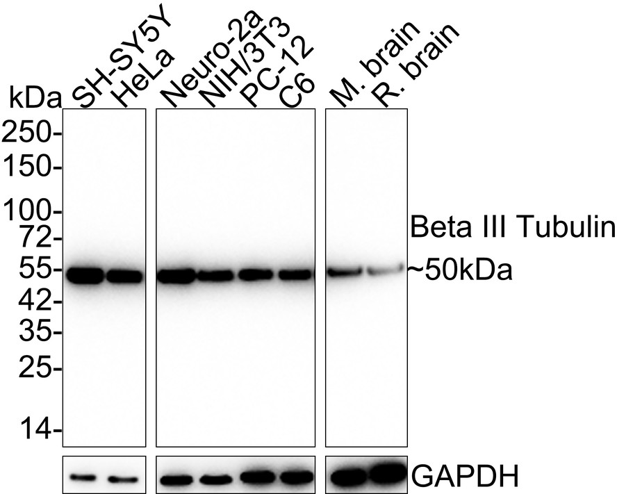
WB
Western blot analysis of Beta III Tubulin on different lysates with Rabbit anti-Beta III Tubulin antibody at 1/20,000 dilution. Lane 1: SH-SY5Y cell lysate (15 µg/Lane) Lane 2: HeLa cell lysate (15 µg/Lane) Lane 3: Neuro-2a cell lysate (15 µg/Lane) Lane 4: NIH/3T3 cell lysate (15 µg/Lane) Lane 5: PC-12 cell lysate (15 µg/Lane) Lane 6: C6 cell lysate (15 µg/Lane) Lane 7: Mouse brain tissue lysate (20 µg/Lane) Lane 8: Rat brain tissue lysate (20 µg/Lane) Exposure time: 3 minutes 54 seconds; 4-20% SDS-PAGE gel. Proteins were transferred to a PVDF membrane and blocked with 5% NFDM/TBST for 1 hour at room temperature. The primary antibody at 1/20,000 dilution was used in 5% NFDM/TBST at room temperature for 2 hours. Goat Anti-Rabbit IgG - HRP Secondary Antibody at 1:50,000 dilution was used for 1 hour at room temperature.IHC
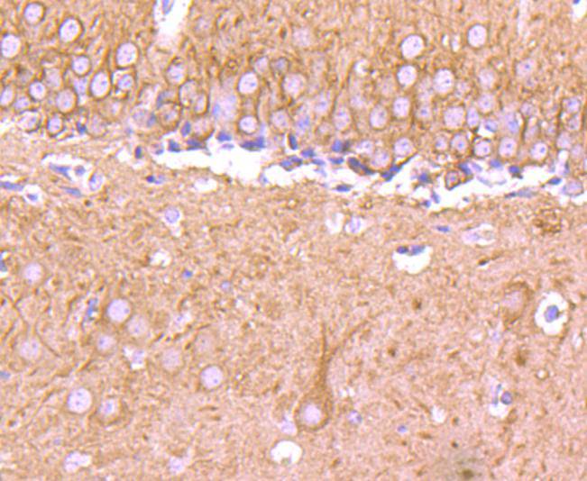
Immunohistochemical analysis of paraffin-embedded mouse brain tissue using anti-Beta III Tubulin antibody. The section was pre-treated using heat mediated antigen retrieval with Tris-EDTA buffer (pH 8.0-8.4) for 20 minutes.The tissues were blocked in 5% BSA for 30 minutes at room temperature, washed with ddH2O and PBS, and then probed with the primary antibody (1/50) for 30 minutes at room temperature. The detection was performed using an HRP conjugated compact polymer system. DAB was used as the chromogen. Tissues were counterstained with hematoxylin and mounted with DPX.ICC/IF
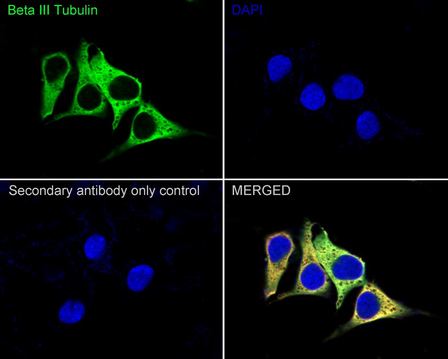
Immunocytochemistry analysis of SH-SY5Y cells labeling Beta III Tubulin with Rabbit anti-Beta III Tubulin antibody at 1/100 dilution. Cells were fixed in 4% paraformaldehyde for 20 minutes at room temperature, permeabilized with 0.1% Triton X-100 in PBS for 5 minutes at room temperature, then blocked with 1% BSA in 10% negative goat serum for 1 hour at room temperature. Cells were then incubated with Rabbit anti-Beta III Tubulin antibody at 1/100 dilution in 1% BSA in PBST overnight at 4 ℃. Goat Anti-Rabbit IgG H&L (iFluor™ 488) was used as the secondary antibody at 1/1,000 dilution. PBS instead of the primary antibody was used as the secondary antibody only control. Nuclear DNA was labelled in blue with DAPI. Beta tubulin (red) was stained at 1/100 dilution overnight at +4℃. Goat Anti-Mouse IgG H&L (iFluor™ 594) was used as the secondary antibody at 1/1,000 dilution.IF-F
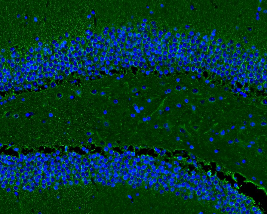
Immunofluorescence analysis of frozen mouse hippocampus (DG) tissue labeling Beta III Tubulin with Rabbit anti-Beta III Tubulin antibody. The tissues were blocked in 3% BSA for 30 minutes at room temperature, washed with PBS, and then probed with the primary antibody (green) at 1/100 dilution overnight at 4℃, washed with PBS. Goat Anti-Rabbit IgG H&L (Alexa Fluor® 488) was used as the secondary antibody at 1/200 dilution. Nuclei were counterstained with DAPI (blue). Image acquisition was performed with KFBIO KF-FL-400 Scanner.FC
Flow cytometric analysis of SH-SY5Y cells labeling Beta III Tubulin. Cells were fixed and permeabilized. Then stained with the primary antibody (1/100) (red) compared with Rabbit IgG Isotype Control (green). After incubation of the primary antibody at +4℃ for an hour, the cells were stained with a iFluor™ 488 conjugate-Goat anti-Rabbit IgG Secondary antibody at 1/1,000 dilution for 30 minutes at +4℃. Unlabelled sample was used as a control (cells without incubation with primary antibody; black).IP
Beta III Tubulin was immunoprecipitated from 0.2 mg SH-SY5Y cell lysate with Rabbit anti-Beta III Tubulin antibody at 2 µg/25 µl agarose. Western blot was performed from the immunoprecipitate using Rabbit anti-Beta III Tubulin antibody at 1/10,000 dilution. Anti-Rabbit IgG for IP Nano-secondary antibody at 1/5,000 dilution was used for 1 hour at room temperature. Lane 1: SH-SY5Y cell lysate (input), Lane 2:Rabbit anti-Beta III Tubulin antibody IP in SH-SY5Y cell lysate, Lane 3: Rabbit IgG instead of Rabbit anti-Beta III Tubulin antibody in SH-SY5Y cell lysate. Blocking/Dilution buffer: 5% NFDM/TBST, Exposure time: 2 seconds.| Product Name | Beta III Tubulin Recombinant Rabbit Monoclonal Antibody |
|---|---|
| Antibody Type | Primary Antibodies |
| Immunogen | Synthetic peptide within Human Tubulin beta-III aa 392-441 / 450. |
| Clonality | Monoclonal |
|---|---|
| Isotype | IgG |
| Host Species | Rabbit |
| Tested Applications | FCICC/IFIF-FIHCIPWB |
| WB:1:20000 IHC:1:50-1:200 ICC:1:100-1:200 IF-F:1:100 FC:1:100 IP:1-2μg/sample |
|
| Species Reactivity | HumanMouseRat |
| Concentration | 1mg/ml |
| Purification | Protein A |
| Gene Symbol | TUBB3 |
|---|---|
| Gene Synonyms | CDCBM FEOM3 TUBB4 CDCBM1 CFEOM3 beta-4 CFEOM3A |
| Gene Full Name | tubulin beta 3 class III |
| Gene Summary | This gene encodes a class III member of the beta tubulin protein family. Beta tubulins are one of two core protein families (alpha and beta tubulins) that heterodimerize and assemble to form microtubules. This protein is primarily expressed in neurons and may be involved in neurogenesis and axon guidance and maintenance. Mutations in this gene are the cause of congenital fibrosis of the extraocular muscles type 3. Alternate splicing results in multiple transcript variants. A pseudogene of this gene is found on chromosome 6. [provided by RefSeq, Oct 2010] |
| Molecular Weight(MW) | 50kDa |
| Cellular Localization | Cytoplasm. |
WB
Western blot analysis of Beta III Tubulin on different lysates with Rabbit anti-Beta III Tubulin antibody at 1/20,000 dilution. Lane 1: SH-SY5Y cell lysate (15 µg/Lane) Lane 2: HeLa cell lysate (15 µg/Lane) Lane 3: Neuro-2a cell lysate (15 µg/Lane) Lane 4: NIH/3T3 cell lysate (15 µg/Lane) Lane 5: PC-12 cell lysate (15 µg/Lane) Lane 6: C6 cell lysate (15 µg/Lane) Lane 7: Mouse brain tissue lysate (20 µg/Lane) Lane 8: Rat brain tissue lysate (20 µg/Lane) Exposure time: 3 minutes 54 seconds; 4-20% SDS-PAGE gel. Proteins were transferred to a PVDF membrane and blocked with 5% NFDM/TBST for 1 hour at room temperature. The primary antibody at 1/20,000 dilution was used in 5% NFDM/TBST at room temperature for 2 hours. Goat Anti-Rabbit IgG - HRP Secondary Antibody at 1:50,000 dilution was used for 1 hour at room temperature.
IHC
Immunohistochemical analysis of paraffin-embedded mouse brain tissue using anti-Beta III Tubulin antibody. The section was pre-treated using heat mediated antigen retrieval with Tris-EDTA buffer (pH 8.0-8.4) for 20 minutes.The tissues were blocked in 5% BSA for 30 minutes at room temperature, washed with ddH2O and PBS, and then probed with the primary antibody (1/50) for 30 minutes at room temperature. The detection was performed using an HRP conjugated compact polymer system. DAB was used as the chromogen. Tissues were counterstained with hematoxylin and mounted with DPX.
ICC/IF
Immunocytochemistry analysis of SH-SY5Y cells labeling Beta III Tubulin with Rabbit anti-Beta III Tubulin antibody at 1/100 dilution. Cells were fixed in 4% paraformaldehyde for 20 minutes at room temperature, permeabilized with 0.1% Triton X-100 in PBS for 5 minutes at room temperature, then blocked with 1% BSA in 10% negative goat serum for 1 hour at room temperature. Cells were then incubated with Rabbit anti-Beta III Tubulin antibody at 1/100 dilution in 1% BSA in PBST overnight at 4 ℃. Goat Anti-Rabbit IgG H&L (iFluor™ 488) was used as the secondary antibody at 1/1,000 dilution. PBS instead of the primary antibody was used as the secondary antibody only control. Nuclear DNA was labelled in blue with DAPI. Beta tubulin (red) was stained at 1/100 dilution overnight at +4℃. Goat Anti-Mouse IgG H&L (iFluor™ 594) was used as the secondary antibody at 1/1,000 dilution.
IF-F
Immunofluorescence analysis of frozen mouse hippocampus (DG) tissue labeling Beta III Tubulin with Rabbit anti-Beta III Tubulin antibody. The tissues were blocked in 3% BSA for 30 minutes at room temperature, washed with PBS, and then probed with the primary antibody (green) at 1/100 dilution overnight at 4℃, washed with PBS. Goat Anti-Rabbit IgG H&L (Alexa Fluor® 488) was used as the secondary antibody at 1/200 dilution. Nuclei were counterstained with DAPI (blue). Image acquisition was performed with KFBIO KF-FL-400 Scanner.
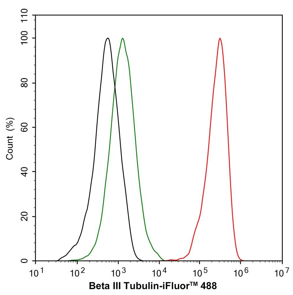
FC
Flow cytometric analysis of SH-SY5Y cells labeling Beta III Tubulin. Cells were fixed and permeabilized. Then stained with the primary antibody (1/100) (red) compared with Rabbit IgG Isotype Control (green). After incubation of the primary antibody at +4℃ for an hour, the cells were stained with a iFluor™ 488 conjugate-Goat anti-Rabbit IgG Secondary antibody at 1/1,000 dilution for 30 minutes at +4℃. Unlabelled sample was used as a control (cells without incubation with primary antibody; black).
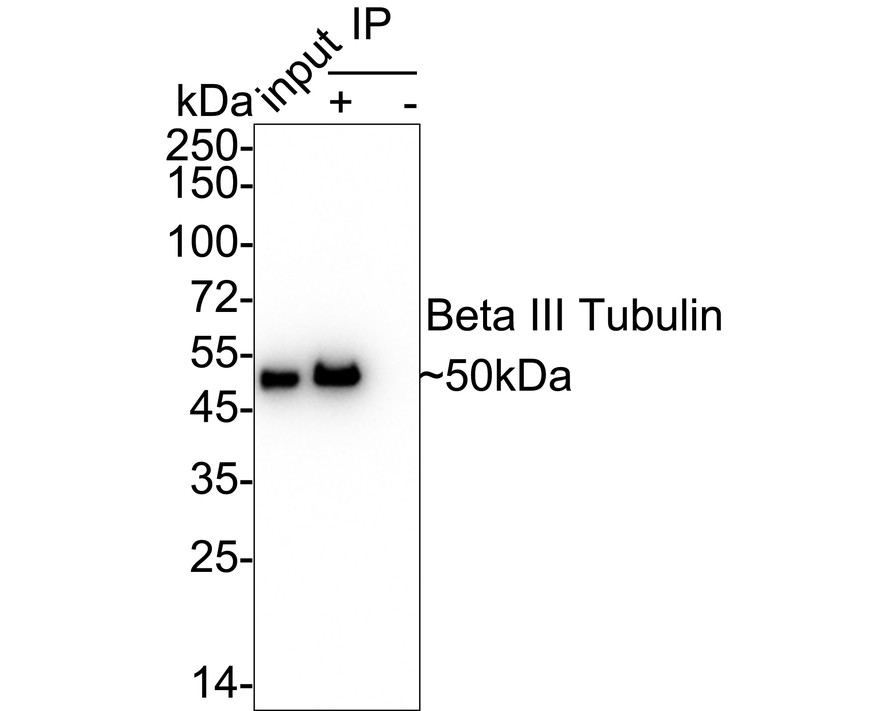
IP
Beta III Tubulin was immunoprecipitated from 0.2 mg SH-SY5Y cell lysate with Rabbit anti-Beta III Tubulin antibody at 2 µg/25 µl agarose. Western blot was performed from the immunoprecipitate using Rabbit anti-Beta III Tubulin antibody at 1/10,000 dilution. Anti-Rabbit IgG for IP Nano-secondary antibody at 1/5,000 dilution was used for 1 hour at room temperature. Lane 1: SH-SY5Y cell lysate (input), Lane 2:Rabbit anti-Beta III Tubulin antibody IP in SH-SY5Y cell lysate, Lane 3: Rabbit IgG instead of Rabbit anti-Beta III Tubulin antibody in SH-SY5Y cell lysate. Blocking/Dilution buffer: 5% NFDM/TBST, Exposure time: 2 seconds.| Application Notes | WB:1:20000 IHC:1:50-1:200 ICC:1:100-1:200 IF-F:1:100 FC:1:100 IP:1-2μg/sample |
|---|
| Form | Liquid |
|---|---|
| Storage Instructions | Store at +4℃ after thawing. Aliquot store at -20℃ or -80℃. Avoid repeated freeze / thaw cycles |
| Storage Buffer | 1*TBS (pH7.4), 0.05% BSA, 40% Glycerol. Preservative: 0.05% Sodium Azide. |


 Data sheet for OM643553