


WB
Various lysates were subjected to SDS PAGE followed by western blot with RORA antibody at dilution of 1:1500 incubated at room temperature for 1.5 hours.IHC
Immunohistochemical analysis of paraffin embedded rat cerebellum tissue slide using RORA antibody at dilution of 1:200 (under 40x lens). Heat mediated antigen retrieval with Tris-EDTA buffer (pH 9.0).ICC/IF
Immunofluorescent analysis of (4% PFA) fixed HepG2 cells using RORA antibody at dilution of 1:300 and 488-Conjugated AffiniPure Goat Anti Rabbit IgG(H+L), CL594-Phalloidin (red).IF-P
Immunofluorescent analysis of (4% PFA) fixed paraffin-embedded rat brain tissue using RORA antibody at dilution of 1:350 and Multi-rAb 488-Goat Anti-Rabbit Recombinant Secondary Antibody (H+L). Heat mediated antigen retrieval with Tris-EDTA buffer (pH 9.0).FC
1x10^6 HeLa cells were intracellularly stained with 0.4 ug RORA Recombinant antibody and 488-Conjugated AffiniPure Goat Anti-Rabbit IgG(H+L)(red), or 0.4 ug Isotype Control (blue). Cells were fixed and permeabilized with Transcription Factor Staining Buffer Kit.WB
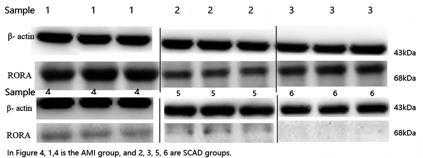
Western blot of some samples from each group. Relative expression of RORA was based on actual calculation, and the internal reference protein and the target protein were compared at the same time. Each specimen was measured three times. Samples 1 and 4 were from the AMI group, and samples 2, 3, 5, and 6 were from the SCAD group.| Product Name | RORA Recombinant antibody |
|---|---|
| Antibody Type | Primary Antibodies |
| Immunogen | Recombinant human RORA protein |
| Clonality | Monoclonal |
|---|---|
| Isotype | IgG |
| Host Species | Rabbit |
| Tested Applications | FCICC/IFIF-PIHCWB |
| WB:1:500-1:3000 IHC:1:50-1:500 ICC:1:150-1:600 IF-P:1:175-1:700 FC:0.40 ug per 10^6 cells in a 100 µl suspension |
|
| Species Reactivity | HumanMouseRat |
| Concentration | 0.3mg/ml |
| Purification | Protein A |
| Gene Symbol | RORA |
|---|---|
| Gene Synonyms | ROR1 ROR2 ROR3 RZRA NR1F1 RORa1 IDDECA RORalpha RZR-ALPHA |
| Gene Full Name | RAR related orphan receptor A |
| Gene Summary | The protein encoded by this gene is a member of the NR1 subfamily of nuclear hormone receptors. It can bind as a monomer or as a homodimer to hormone response elements upstream of several genes to enhance the expression of those genes. The encoded protein has been shown to interact with NM23-2, a nucleoside diphosphate kinase involved in organogenesis and differentiation, as well as with NM23-1, the product of a tumor metastasis suppressor candidate gene. Also, it has been shown to aid in the transcriptional regulation of some genes involved in circadian rhythm. Four transcript variants encoding different isoforms have been described for this gene. [provided by RefSeq, Feb 2014] |
| Alternative Names | 230217G6 NR1F1 Nuclear receptor ROR-alpha Nuclear receptor RZR-alpha RAR-related orphan receptor A |
| Molecular Weight(MW) | 59kDa(Observed Molecular Weight 60-66kDa) |
| Cellular Localization | Nucleus. |
WB
Various lysates were subjected to SDS PAGE followed by western blot with RORA antibody at dilution of 1:1500 incubated at room temperature for 1.5 hours.
IHC
Immunohistochemical analysis of paraffin embedded rat cerebellum tissue slide using RORA antibody at dilution of 1:200 (under 40x lens). Heat mediated antigen retrieval with Tris-EDTA buffer (pH 9.0).
ICC/IF
Immunofluorescent analysis of (4% PFA) fixed HepG2 cells using RORA antibody at dilution of 1:300 and 488-Conjugated AffiniPure Goat Anti Rabbit IgG(H+L), CL594-Phalloidin (red).
IF-P
Immunofluorescent analysis of (4% PFA) fixed paraffin-embedded rat brain tissue using RORA antibody at dilution of 1:350 and Multi-rAb 488-Goat Anti-Rabbit Recombinant Secondary Antibody (H+L). Heat mediated antigen retrieval with Tris-EDTA buffer (pH 9.0).
FC
1x10^6 HeLa cells were intracellularly stained with 0.4 ug RORA Recombinant antibody and 488-Conjugated AffiniPure Goat Anti-Rabbit IgG(H+L)(red), or 0.4 ug Isotype Control (blue). Cells were fixed and permeabilized with Transcription Factor Staining Buffer Kit.
WB
Western blot of some samples from each group. Relative expression of RORA was based on actual calculation, and the internal reference protein and the target protein were compared at the same time. Each specimen was measured three times. Samples 1 and 4 were from the AMI group, and samples 2, 3, 5, and 6 were from the SCAD group.| Application Notes | WB:1:500-1:3000 IHC:1:50-1:500 ICC:1:150-1:600 IF-P:1:175-1:700 FC:0.40 ug per 10^6 cells in a 100 µl suspension |
|---|
| Form | Liquid |
|---|---|
| Storage Instructions | Store at -20°C. Stable for one year after shipment. |
| Storage Buffer | PBS with 0.02% sodium azide and 50% glycerol pH 7.3. |


