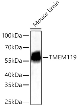

WB
Western blot analysis of lysates from Mouse brain using TMEM119 Rabbit mAb at 1:12000 dilution incubated overnight at 4℃. Secondary antibody: HRP-conjugated Goat anti-Rabbit IgG(H+L) at 1:10000 dilution. Lysates/proteins: 25 μg per lane. Blocking buffer: 3% nonfat dry milk in TBST. Detection: ECL Basic Kit. Exposure time: 45s.IHC
Immunohistochemistry analysis of paraffin embedded Mouse intestine tissue using TMEM119 Rabbit mAb at a dilution of 1:6000 (40x lens). High pressure antigen retrieval performed with 0.01M Tris-EDTA Buffer (pH 9.0) prior to IHC staining.IF-P
Confocal imaging of paraffin-embedded Mouse brain tissue using TMEM119 Rabbit mAb (dilution 1:500) followed by a further incubation with Cy3 Goat Anti-Rabbit IgG (H+L) (dilution 1:500) (Red). DAPI was used for nuclear staining (Blue). High pressure antigen retrieval performed with 0.01M Citrate Buffer (pH 6.0) prior to IF staining. Objective: 40x.| Product Name | TMEM119 Rabbit mAb |
|---|---|
| Antibody Type | Primary Antibodies |
| Immunogen | Recombinant protein of human TMEM119. |
| Clonality | monoclonal |
|---|---|
| Isotype | IgG |
| Host Species | Rabbit |
| Tested Applications | IF-PIHCWB |
| WB:1:6000-1:24000 IHC:1:2000-1:8000 IF-P:1:500-1:2000 |
|
| Species Reactivity | Mouse |
| Concentration | 1mg/ml |
| Purification | Affinity purified |
| Gene Symbol | TMEM119 |
|---|---|
| Gene Synonyms | OBIF |
| Gene Full Name | transmembrane protein 119 |
| Gene Summary | Involved in positive regulation of bone mineralization; positive regulation of osteoblast differentiation; and positive regulation of osteoblast proliferation. Located in plasma membrane. [provided by Alliance of Genome Resources, Feb 2025] |
| Molecular Weight(MW) | 29kDa(Observed MW 56kDa) |
| Cellular Localization | plasma membrane. |
WB
Western blot analysis of lysates from Mouse brain using TMEM119 Rabbit mAb at 1:12000 dilution incubated overnight at 4℃. Secondary antibody: HRP-conjugated Goat anti-Rabbit IgG(H+L) at 1:10000 dilution. Lysates/proteins: 25 μg per lane. Blocking buffer: 3% nonfat dry milk in TBST. Detection: ECL Basic Kit. Exposure time: 45s.
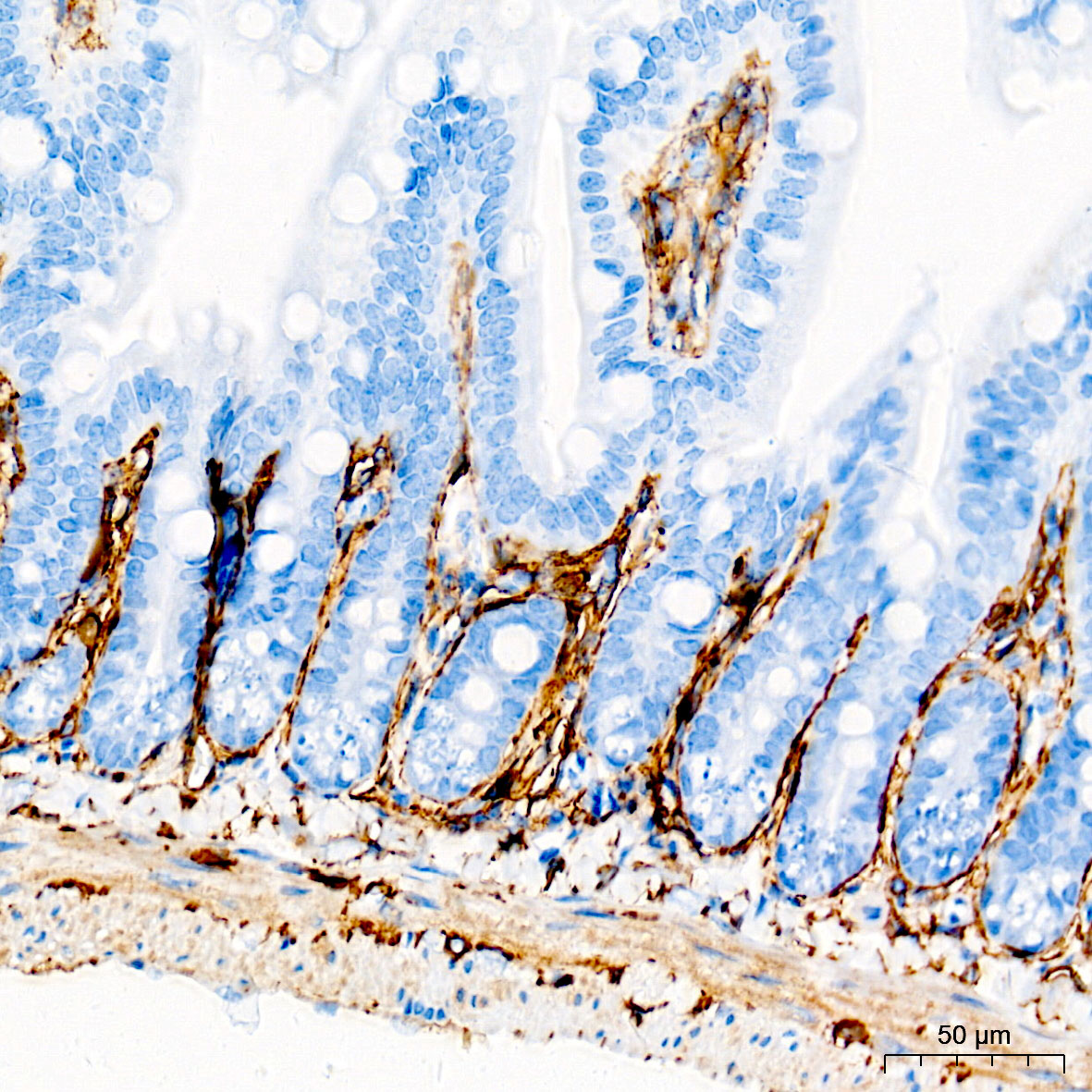
IHC
Immunohistochemistry analysis of paraffin embedded Mouse intestine tissue using TMEM119 Rabbit mAb at a dilution of 1:6000 (40x lens). High pressure antigen retrieval performed with 0.01M Tris-EDTA Buffer (pH 9.0) prior to IHC staining.
IF-P
Confocal imaging of paraffin-embedded Mouse brain tissue using TMEM119 Rabbit mAb (dilution 1:500) followed by a further incubation with Cy3 Goat Anti-Rabbit IgG (H+L) (dilution 1:500) (Red). DAPI was used for nuclear staining (Blue). High pressure antigen retrieval performed with 0.01M Citrate Buffer (pH 6.0) prior to IF staining. Objective: 40x.| Application Notes | WB:1:6000-1:24000 IHC:1:2000-1:8000 IF-P:1:500-1:2000 |
|---|
| Form | Liquid |
|---|---|
| Storage Instructions | Store at -20℃. Avoid freeze / thaw cycles. |
| Storage Buffer | Buffer: PBS with 0.05% proclin300, 0.05% BSA, 50% glycerol, pH7.3. |


 Data sheet for OM643856