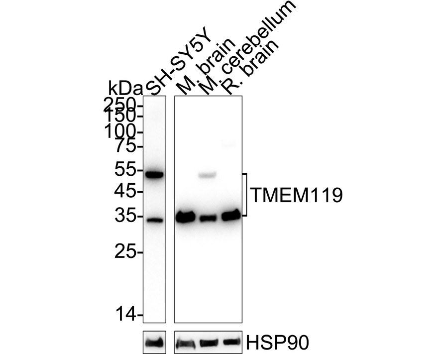
WB
Western blot analysis of TMEM119 on different lysates with Rabbit anti-TMEM119 antibody at 1/2,000 dilution. Lane 1: SH-SY5Y cell lysate (20 µg/Lane), Lane 2: Mouse brain tissue lysate (40 µg/Lane), Lane 3: Mouse cerebellum tissue lysate (40 µg/Lane), Lane 4: Rat brain tissue lysate (40 µg/Lane), Exposure time: Lane 1: 3 minutes; Lane 2-4: 14 seconds; 4-20% SDS-PAGE gel. Proteins were transferred to a PVDF membrane and blocked with 5% NFDM/TBST for 1 hour at room temperature. The primary antibody at 1/2,000 dilution was used in 5% NFDM/TBST at 4℃ overnight. Goat Anti-Rabbit IgG - HRP Secondary Antibody at 1/50,000 dilution was used for 1 hour at room temperature.ICC/IF
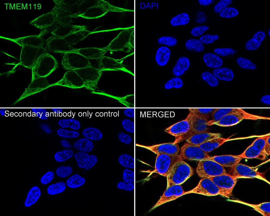
Immunocytochemistry analysis of SH-SY5Y cells labeling TMEM119 with Rabbit anti-TMEM119 antibody at 1/200 dilution. Cells were fixed in 4% paraformaldehyde for 15 minutes at room temperature, permeabilized with 0.1% Triton X-100 in PBS for 15 minutes at room temperature, then blocked with 1% BSA in 10% negative goat serum for 1 hour at room temperature. Cells were then incubated with Rabbit anti-TMEM119 antibody at 1/200 dilution in 1% BSA in PBST overnight at 4 ℃. Goat Anti-Rabbit IgG H&L (488) was used as the secondary antibody at 1/1,000 dilution. PBS instead of the primary antibody was used as the secondary antibody only control. Nuclear DNA was labelled in blue with DAPI. Beta tubulin (red) was stained at 1/100 dilution overnight at +4℃. Goat Anti-Mouse IgG H&L (594) was used as the secondary antibody at 1/1,000 dilution.FC
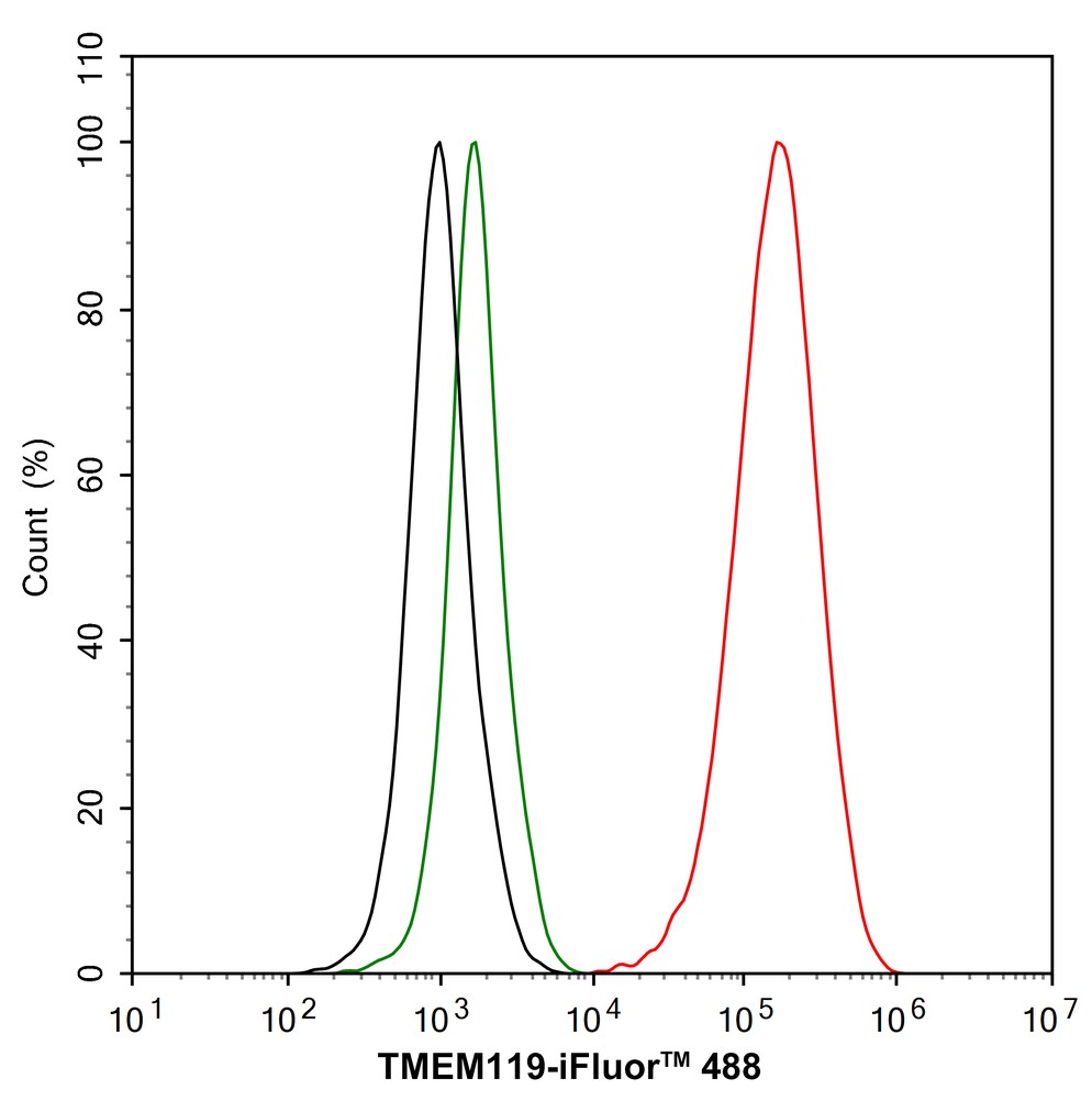
Flow cytometric analysis of SH-SY5Y cells labeling TMEM119. Cells were fixed and permeabilized. Then stained with the primary antibody (1/1,000) (red) compared with Rabbit IgG Isotype Control (green). After incubation of the primary antibody at +4℃ for an hour, the cells were stained with a 488 conjugate-Goat anti-Rabbit IgG Secondary antibody at 1/1,000 dilution for 30 minutes at +4℃. Unlabelled sample was used as a control (cells without incubation with primary antibody; black).IP
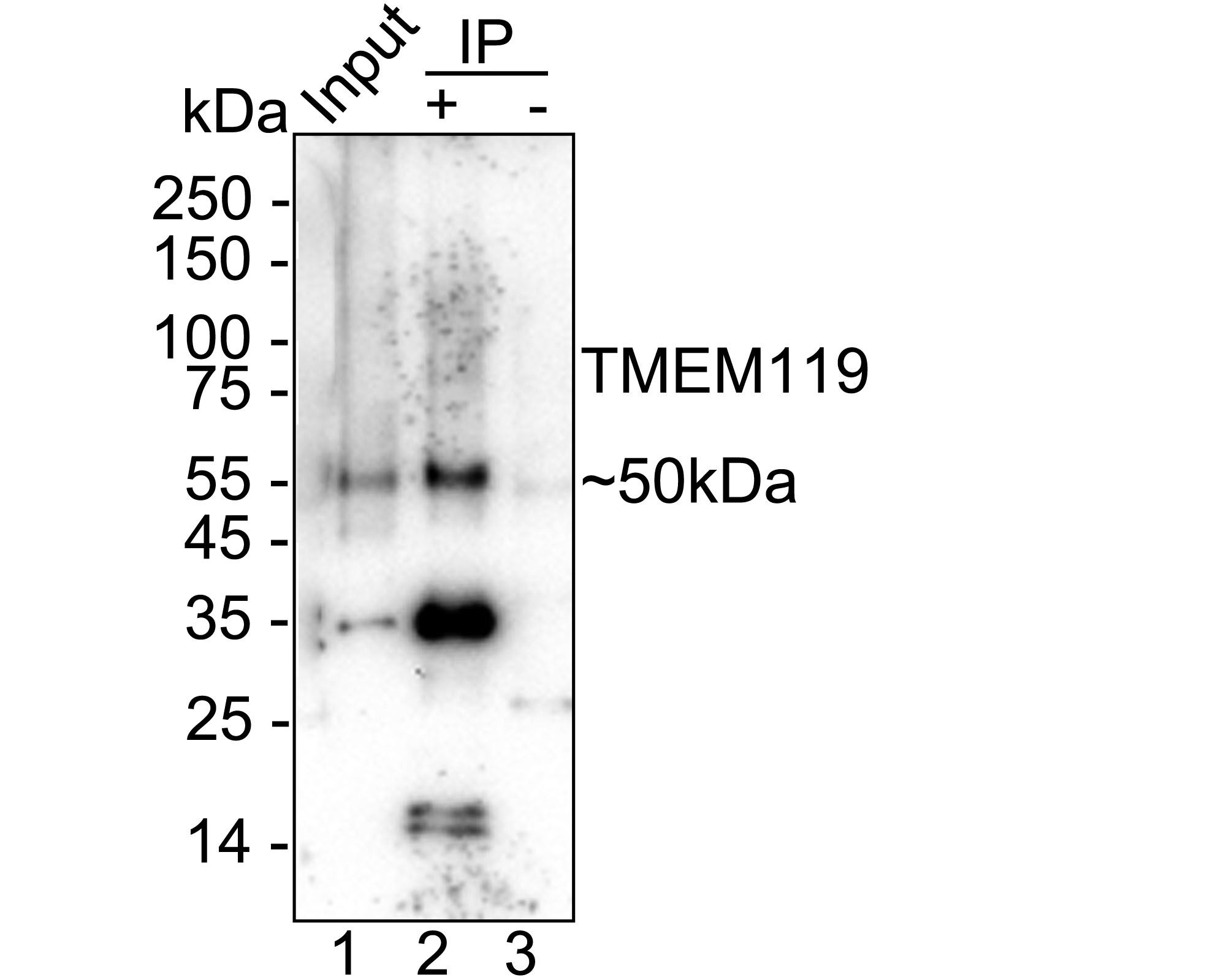
TMEM119 was immunoprecipitated from 0.2 mg mouse cerebellum tissue lysate with Rabbit anti-TMEM119 antibody at 2µg/10µl beads. Western blot was performed from the immunoprecipitate using Rabbit anti-TMEM119 antibody at 1/5,000 dilution. HRP Conjugated Anti-Rabbit IgG for IP Nano-secondary antibody at 1/5,000 dilution was used for 1 hour at room temperature. Lane 1: Mouse cerebellum tissue lysate (input), Lane 2: Rabbit anti-TMEM119 antibody IP in mouse cerebellum tissue lysate, Lane 3: Rabbit IgG instead of Rabbit anti-TMEM119 antibody in mouse cerebellum tissue lysate, Blocking/Dilution buffer: primary antibody dilution. Exposure time: 3 minutes.| Product Name | TMEM119 Recombinant Rabbit Monoclonal Antibody |
|---|---|
| Antibody Type | Primary Antibodies |
| Immunogen | Recombinant protein within mouse TMEM119 aa 113-280. |
| Clonality | monoclonal |
|---|---|
| Isotype | IgG |
| Host Species | Rabbit |
| Tested Applications | FCICC/IFIPWB |
| WB:1:2000 ICC/IF:1:100 FC:1:1000 IP:1-2μg/sample |
|
| Species Reactivity | HumanMouseRat |
| Concentration | 1mg/ml |
| Purification | Protein A |
| Gene Symbol | TMEM119 |
|---|---|
| Gene Synonyms | OBIF |
| Gene Full Name | transmembrane protein 119 |
| Gene Summary | Involved in positive regulation of bone mineralization; positive regulation of osteoblast differentiation; and positive regulation of osteoblast proliferation. Located in plasma membrane. [provided by Alliance of Genome Resources, Apr 2025] |
| Molecular Weight(MW) | 29kDa(Observed band size: 35/55kDa) |
| Cellular Localization | Cell membrane, Cytoplasm, Endoplasmic reticulum membrane, Secreted. |
WB
Western blot analysis of TMEM119 on different lysates with Rabbit anti-TMEM119 antibody at 1/2,000 dilution. Lane 1: SH-SY5Y cell lysate (20 µg/Lane), Lane 2: Mouse brain tissue lysate (40 µg/Lane), Lane 3: Mouse cerebellum tissue lysate (40 µg/Lane), Lane 4: Rat brain tissue lysate (40 µg/Lane), Exposure time: Lane 1: 3 minutes; Lane 2-4: 14 seconds; 4-20% SDS-PAGE gel. Proteins were transferred to a PVDF membrane and blocked with 5% NFDM/TBST for 1 hour at room temperature. The primary antibody at 1/2,000 dilution was used in 5% NFDM/TBST at 4℃ overnight. Goat Anti-Rabbit IgG - HRP Secondary Antibody at 1/50,000 dilution was used for 1 hour at room temperature.
ICC/IF
Immunocytochemistry analysis of SH-SY5Y cells labeling TMEM119 with Rabbit anti-TMEM119 antibody at 1/200 dilution. Cells were fixed in 4% paraformaldehyde for 15 minutes at room temperature, permeabilized with 0.1% Triton X-100 in PBS for 15 minutes at room temperature, then blocked with 1% BSA in 10% negative goat serum for 1 hour at room temperature. Cells were then incubated with Rabbit anti-TMEM119 antibody at 1/200 dilution in 1% BSA in PBST overnight at 4 ℃. Goat Anti-Rabbit IgG H&L (488) was used as the secondary antibody at 1/1,000 dilution. PBS instead of the primary antibody was used as the secondary antibody only control. Nuclear DNA was labelled in blue with DAPI. Beta tubulin (red) was stained at 1/100 dilution overnight at +4℃. Goat Anti-Mouse IgG H&L (594) was used as the secondary antibody at 1/1,000 dilution.
FC
Flow cytometric analysis of SH-SY5Y cells labeling TMEM119. Cells were fixed and permeabilized. Then stained with the primary antibody (1/1,000) (red) compared with Rabbit IgG Isotype Control (green). After incubation of the primary antibody at +4℃ for an hour, the cells were stained with a 488 conjugate-Goat anti-Rabbit IgG Secondary antibody at 1/1,000 dilution for 30 minutes at +4℃. Unlabelled sample was used as a control (cells without incubation with primary antibody; black).
IP
TMEM119 was immunoprecipitated from 0.2 mg mouse cerebellum tissue lysate with Rabbit anti-TMEM119 antibody at 2µg/10µl beads. Western blot was performed from the immunoprecipitate using Rabbit anti-TMEM119 antibody at 1/5,000 dilution. HRP Conjugated Anti-Rabbit IgG for IP Nano-secondary antibody at 1/5,000 dilution was used for 1 hour at room temperature. Lane 1: Mouse cerebellum tissue lysate (input), Lane 2: Rabbit anti-TMEM119 antibody IP in mouse cerebellum tissue lysate, Lane 3: Rabbit IgG instead of Rabbit anti-TMEM119 antibody in mouse cerebellum tissue lysate, Blocking/Dilution buffer: primary antibody dilution. Exposure time: 3 minutes.| Application Notes | WB:1:2000 ICC/IF:1:100 FC:1:1000 IP:1-2μg/sample |
|---|
| Form | Liquid |
|---|---|
| Storage Instructions | Store at +4℃ after thawing. Aliquot store at -20℃. Avoid repeated freeze / thaw cycles. |
| Storage Buffer | PBS (pH7.4), 0.1% BSA, 40% Glycerol. Preservative: 0.05% Sodium Azide. |


 Data sheet for OM644235