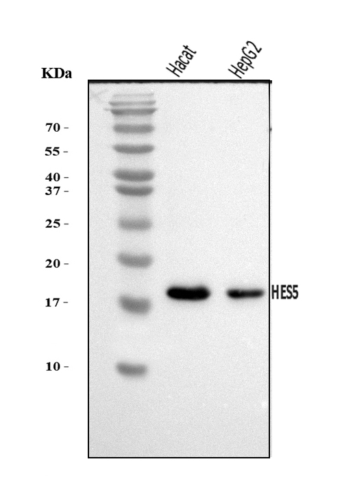
WB
Western blot analysis of HES5 using anti-HES5 antibody. The sample well of each lane was loaded with 30 ug of sample under reducing conditions. Lane 1: Hacat whole cell lysates, Lane 2: HepG2 whole cell lysates. After electrophoresis, proteins were transferred to a membrane. Then the membrane was incubated with rabbit anti-HES5 antigen affinity purified polyclonal antibody at a dilution of 1:1000 and probed with a goat anti-rabbit IgG-HRP secondary antibody. The signal is developed using ECL Plus Western Blotting Substrate.IHC
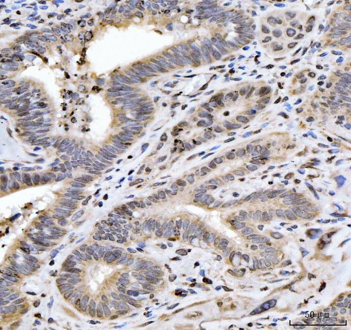
IHC analysis of HES5 using anti-HES5 antibody. HES5 was detected in a paraffin-embedded section of human Gall bladder adenosquamous carcinoma tissue. Biotinylated goat anti-rabbit IgG was used as secondary antibody. The tissue section was incubated with rabbit anti-HES5 Antibody at a dilution of 1:200 and developed using Strepavidin-Biotin-Complex (SABC) with DAB as the chromogen.FC
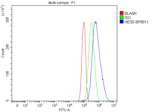
Flow Cytometry analysis of PC-3 cells using anti-HES5 antibody. Overlay histogram showing PC-3 cells stained with anti-HES5 antibody (Blue line). To facilitate intracellular staining, cells were fixed with 4% paraformaldehyde and permeabilized with permeabilization buffer. The cells were blocked with 10% normal goat serum. And then incubated with rabbit anti-HES5 Antibody at 1:100 dilution for 30 min at 20°C. 488 conjugated goat anti-rabbit IgG was used as secondary antibody at 1:100 dilution for 30 minutes at 20°C. Isotype control antibody (Green line) was rabbit IgG at 1:100 dilution used under the same conditions. Unlabelled sample without incubation with primary antibody and secondary antibody (Red line) was used as a blank control.| Product Name | Rabbit polyclonal antibody to HES5 |
|---|---|
| Antibody Type | Primary Antibodies |
| Immunogen | E.coli-derived human HES5 recombinant protein (Position: M1-R121). |
| Clonality | polyclonal |
|---|---|
| Isotype | IgG |
| Host Species | Rabbit |
| Tested Applications | FCIHCWB |
| WB:1:500-1:2000 IHC:1:50-1:400 FC:1:50-1:200 |
|
| Species Reactivity | Human |
| Concentration | 0.5mg/ml |
| Purification | Affinity purified |
| Gene Symbol | HES5 |
|---|---|
| Gene Synonyms | bHLHb38 |
| Gene Full Name | hes family bHLH transcription factor 5 |
| Gene Summary | This gene encodes a member of a family of basic helix-loop-helix transcriptional repressors. The protein product of this gene, which is activated downstream of the Notch pathway, regulates cell differentiation in multiple tissues. Disruptions in the normal expression of this gene have been associated with developmental diseases and cancer. [provided by RefSeq, Dec 2008] |
| Molecular Weight(MW) | 18kDa |
| Cellular Localization | Nucleus. |
WB
Western blot analysis of HES5 using anti-HES5 antibody. The sample well of each lane was loaded with 30 ug of sample under reducing conditions. Lane 1: Hacat whole cell lysates, Lane 2: HepG2 whole cell lysates. After electrophoresis, proteins were transferred to a membrane. Then the membrane was incubated with rabbit anti-HES5 antigen affinity purified polyclonal antibody at a dilution of 1:1000 and probed with a goat anti-rabbit IgG-HRP secondary antibody. The signal is developed using ECL Plus Western Blotting Substrate.
IHC
IHC analysis of HES5 using anti-HES5 antibody. HES5 was detected in a paraffin-embedded section of human Gall bladder adenosquamous carcinoma tissue. Biotinylated goat anti-rabbit IgG was used as secondary antibody. The tissue section was incubated with rabbit anti-HES5 Antibody at a dilution of 1:200 and developed using Strepavidin-Biotin-Complex (SABC) with DAB as the chromogen.
FC
Flow Cytometry analysis of PC-3 cells using anti-HES5 antibody. Overlay histogram showing PC-3 cells stained with anti-HES5 antibody (Blue line). To facilitate intracellular staining, cells were fixed with 4% paraformaldehyde and permeabilized with permeabilization buffer. The cells were blocked with 10% normal goat serum. And then incubated with rabbit anti-HES5 Antibody at 1:100 dilution for 30 min at 20°C. 488 conjugated goat anti-rabbit IgG was used as secondary antibody at 1:100 dilution for 30 minutes at 20°C. Isotype control antibody (Green line) was rabbit IgG at 1:100 dilution used under the same conditions. Unlabelled sample without incubation with primary antibody and secondary antibody (Red line) was used as a blank control.| Application Notes | WB:1:500-1:2000 IHC:1:50-1:400 FC:1:50-1:200 |
|---|
| Form | Liquid |
|---|---|
| Storage Instructions | 12 months from date of receipt, -20℃ as supplied. 6 months 2 to 8℃ after reconstitution. Avoid repeated freezing and thawing. |
| Storage Buffer | 500ug/ml antibody with PBS, 0.02% NaN3, 1 mg/ml BSA and 50% glycerol. |


 Data sheet for OM643937