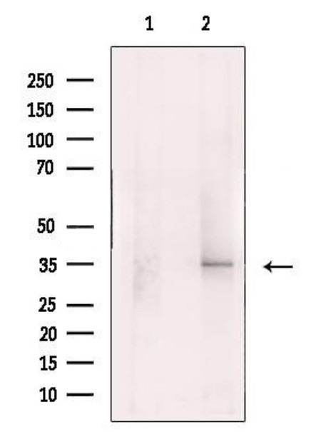
WB
Western blot analysis of extracts from Human spleen, using XAF1 Antibody. The lane on the left was treated with blocking peptide.IHC
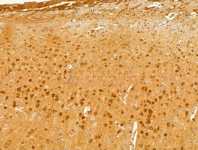
XAF1 Antibody at 1/100 staining Mouse brain tissue by IHC-P. The sample was formaldehyde fixed and a heat mediated antigen retrieval step in citrate buffer was performed. The sample was then blocked and incubated with the primary antibody at 4°C overnight. An HRP conjugated anti-Rabbit antibody was used as the secondary antibody.| Product Name | Rabbit polyclonal antibody to XAF1 |
|---|---|
| Antibody Type | Primary Antibodies |
| Clonality | polyclonal |
|---|---|
| Isotype | IgG |
| Host Species | Rabbit |
| Tested Applications | IHCWB |
| WB:1:500-1:2000 IHC:1:50-1:200 |
|
| Species Reactivity | HumanMouseRat |
| Concentration | 1mg/ml |
| Purification | Affinity purified |
| Gene Symbol | XAF1 |
|---|---|
| Gene Synonyms | BIRC4BP XIAPAF1 HSXIAPAF1 |
| Gene Full Name | XIAP associated factor 1 |
| Gene Summary | This gene encodes a protein which binds to and counteracts the inhibitory effect of a member of the IAP (inhibitor of apoptosis) protein family. IAP proteins bind to and inhibit caspases which are activated during apoptosis. The proportion of IAPs and proteins which interfere with their activity, such as the encoded protein, affect the progress of the apoptosis signaling pathway. Multiple transcript variants encoding different isoforms have been found for this gene. [provided by RefSeq, Feb 2012] |
| Molecular Weight(MW) | 35kDa |
| Cellular Localization | Cytoplasm. Nucleus. Mitochondrion. |
WB
Western blot analysis of extracts from Human spleen, using XAF1 Antibody. The lane on the left was treated with blocking peptide.
IHC
XAF1 Antibody at 1/100 staining Mouse brain tissue by IHC-P. The sample was formaldehyde fixed and a heat mediated antigen retrieval step in citrate buffer was performed. The sample was then blocked and incubated with the primary antibody at 4°C overnight. An HRP conjugated anti-Rabbit antibody was used as the secondary antibody.| Application Notes | WB:1:500-1:2000 IHC:1:50-1:200 |
|---|
| Form | Liquid |
|---|---|
| Storage Instructions | Store at -20 °C. Stable for 12 months from date of receipt. |
| Storage Buffer | Rabbit IgG in phosphate buffered saline , pH 7.4, 150mM NaCl, 0.02% sodium azide and 50% glycerol. |


 Data sheet for OM643993