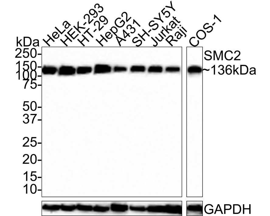

WB
Western blot analysis of SMC2 on different lysates with Rabbit anti-SMC2 antibody at 1/1,000 dilution. Lane 1: HeLa cell lysate (30 µg/Lane), Lane 2: HEK-293 cell lysate (30 µg/Lane), Lane 3: HT-29 cell lysate (30 µg/Lane), Lane 4: HepG2 cell lysate (30 µg/Lane), Lane 5: A431 cell lysate (30 µg/Lane), Lane 6: SH-SY5Y cell lysate (30 µg/Lane), Lane 7: Jurkat cell lysate (30 µg/Lane), Lane 8: Raji cell lysate (30 µg/Lane), Lane 9: COS-1 cell lysate (24 µg/Lane), Exposure time: 30 seconds; 4-20% SDS-PAGE gel. Proteins were transferred to a PVDF membrane and blocked with 5% NFDM/TBST for 1 hour at room temperature. The primary antibody at 1/1,000 dilution was used in 5% NFDM/TBST at room temperature for 2 hours. Goat Anti-Rabbit IgG - HRP Secondary Antibody at 1:100,000 dilution was used for 1 hour at room temperature.IHC
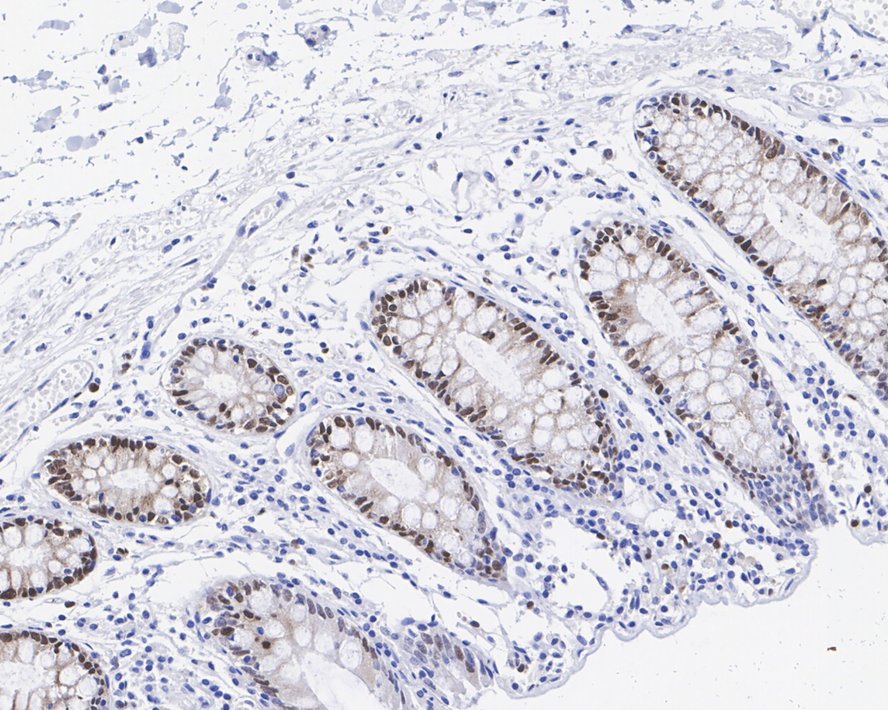
Immunohistochemical analysis of paraffin-embedded human colon tissue with Rabbit anti-SMC2 antibody at 1/1,000 dilution. The section was pre-treated using heat mediated antigen retrieval with sodium citrate buffer (pH 6.0) for 2 minutes. The tissues were blocked in 1% BSA for 20 minutes at room temperature, washed with ddH2O and PBS, and then probed with the primary antibody at 1/1,000 dilution for 1 hour at room temperature. The detection was performed using an HRP conjugated compact polymer system. DAB was used as the chromogen. Tissues were counterstained with hematoxylin and mounted with DPX.ICC/IF
Immunocytochemistry analysis of HeLa cells labeling SMC2 with Rabbit anti-SMC2 antibody at 1/100 dilution. Cells were fixed in 100% methanol for 10 minutes, permeabilized with 0.1% Triton X-100 in PBS for 15 minutes, and then blocked with 1% BSA for 30 minutes at room temperature. Cells were then incubated with Rabbit anti-SMC2 antibody at 1/100 dilution in 2% negative goat serum overnight at 4 ℃. Goat Anti-Rabbit IgG H&L (488) was used as the secondary antibody at 1/1,000 dilution. Nuclear DNA was labelled in blue with DAPI. Beta tubulin (red) was stained at 1/100 dilution overnight at +4℃. Goat Anti-Mouse IgG H&L (iFluor™ 594) was used as the secondary antibody at 1/1,000 dilution.| Product Name | SMC2 Recombinant Rabbit Monoclonal Antibody |
|---|---|
| Antibody Type | Primary Antibodies |
| Immunogen | Recombinant protein within human SMC2 aa 451-750 / 1,197. |
| Clonality | monoclonal |
|---|---|
| Isotype | IgG |
| Host Species | Rabbit |
| Tested Applications | ICC/IFIHCWB |
| WB:1:1000 IHC:1:1000 ICC/IF:1:100 |
|
| Species Reactivity | HumanMouseRat |
| Concentration | 1mg/ml |
| Purification | Protein A |
| Gene Symbol | SMC2 |
|---|---|
| Gene Synonyms | CAPE CAP-E SMC-2 SMC2L1 |
| Gene Full Name | structural maintenance of chromosomes 2 |
| Gene Summary | Predicted to enable chromatin binding activity. Involved in mitotic chromosome condensation and positive regulation of chromosome condensation. Located in condensed chromosome; cytoplasm; and nuclear lumen. Part of condensin complex. Implicated in colon adenocarcinoma. Biomarker of colorectal cancer. [provided by Alliance of Genome Resources, Mar 2025] |
| Molecular Weight(MW) | 136kDa |
| Cellular Localization | Nucleus, Cytoplasm, Chromosome. |
WB
Western blot analysis of SMC2 on different lysates with Rabbit anti-SMC2 antibody at 1/1,000 dilution. Lane 1: HeLa cell lysate (30 µg/Lane), Lane 2: HEK-293 cell lysate (30 µg/Lane), Lane 3: HT-29 cell lysate (30 µg/Lane), Lane 4: HepG2 cell lysate (30 µg/Lane), Lane 5: A431 cell lysate (30 µg/Lane), Lane 6: SH-SY5Y cell lysate (30 µg/Lane), Lane 7: Jurkat cell lysate (30 µg/Lane), Lane 8: Raji cell lysate (30 µg/Lane), Lane 9: COS-1 cell lysate (24 µg/Lane), Exposure time: 30 seconds; 4-20% SDS-PAGE gel. Proteins were transferred to a PVDF membrane and blocked with 5% NFDM/TBST for 1 hour at room temperature. The primary antibody at 1/1,000 dilution was used in 5% NFDM/TBST at room temperature for 2 hours. Goat Anti-Rabbit IgG - HRP Secondary Antibody at 1:100,000 dilution was used for 1 hour at room temperature.
IHC
Immunohistochemical analysis of paraffin-embedded human colon tissue with Rabbit anti-SMC2 antibody at 1/1,000 dilution. The section was pre-treated using heat mediated antigen retrieval with sodium citrate buffer (pH 6.0) for 2 minutes. The tissues were blocked in 1% BSA for 20 minutes at room temperature, washed with ddH2O and PBS, and then probed with the primary antibody at 1/1,000 dilution for 1 hour at room temperature. The detection was performed using an HRP conjugated compact polymer system. DAB was used as the chromogen. Tissues were counterstained with hematoxylin and mounted with DPX.
ICC/IF
Immunocytochemistry analysis of HeLa cells labeling SMC2 with Rabbit anti-SMC2 antibody at 1/100 dilution. Cells were fixed in 100% methanol for 10 minutes, permeabilized with 0.1% Triton X-100 in PBS for 15 minutes, and then blocked with 1% BSA for 30 minutes at room temperature. Cells were then incubated with Rabbit anti-SMC2 antibody at 1/100 dilution in 2% negative goat serum overnight at 4 ℃. Goat Anti-Rabbit IgG H&L (488) was used as the secondary antibody at 1/1,000 dilution. Nuclear DNA was labelled in blue with DAPI. Beta tubulin (red) was stained at 1/100 dilution overnight at +4℃. Goat Anti-Mouse IgG H&L (iFluor™ 594) was used as the secondary antibody at 1/1,000 dilution.| Application Notes | WB:1:1000 IHC:1:1000 ICC/IF:1:100 |
|---|
| Form | Liquid |
|---|---|
| Storage Instructions | Store at +4℃ after thawing. Aliquot store at -20℃. Avoid repeated freeze / thaw cycles. |
| Storage Buffer | 1*TBS (pH7.4), 0.05% BSA, 40% Glycerol. Preservative: 0.05% Sodium Azide. |


 Data sheet for OM644054