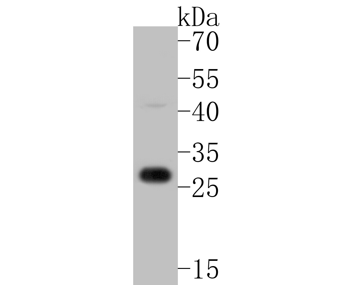
WB
Western blot analysis of Claudin 18.2 on rat heart tissue lysates. Proteins were transferred to a PVDF membrane and blocked with 5% BSA in PBS for 1 hour at room temperature. The primary antibody (1/500) was used in 5% BSA at room temperature for 2 hours. Goat Anti-Rabbit IgG - HRP Secondary Antibody at 1:5,000 dilution was used for 1 hour at room temperature.ICC/IF
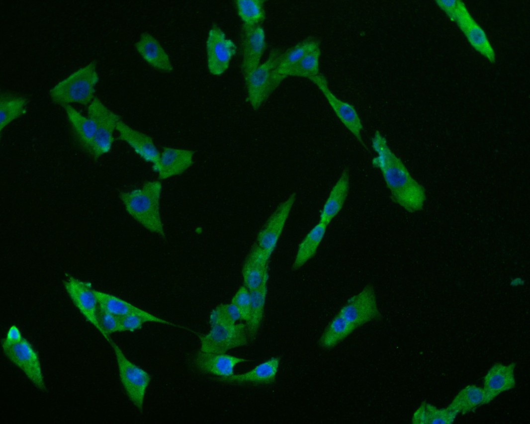
ICC staining of Claudin 18.2 in MG-63 cells (green). Formalin fixed cells were permeabilized with 0.1% Triton X-100 in TBS for 10 minutes at room temperature and blocked with 10% negative goat serum for 15 minutes at room temperature. Cells were probed with the primary antibody (1/50) for 1 hour at room temperature, washed with PBS. Alexa Fluor®488 conjugate-Goat anti-Rabbit IgG was used as the secondary antibody at 1/1,000 dilution. The nuclear counter stain is DAPI (blue).IHC
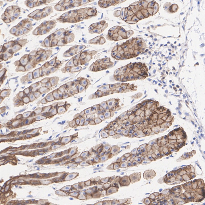
Immunohistochemical analysis of paraffin-embedded mouse stomach tissue with Rabbit anti-Claudin 18.2 antibody at 1/8,000 dilution. The section was pre-treated using heat mediated antigen retrieval with Tris-EDTA buffer (pH 9.0) for 20 minutes. The tissues were blocked in 1% BSA for 20 minutes at room temperature, washed with ddH2O and PBS, and then probed with the primary antibody at 1/8,000 dilution for 1 hour at room temperature. The detection was performed using an HRP conjugated compact polymer system. DAB was used as the chromogen. Tissues were counterstained with hematoxylin and mounted with DPX.FC
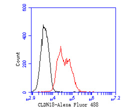
Flow cytometric analysis of Claudin 18.2 was done on F9 cells. The cells were fixed, permeabilized and stained with the primary antibody (1/50) (red). After incubation of the primary antibody at room temperature for an hour, the cells were stained with a Alexa Fluor 488-conjugated Goat anti-Rabbit IgG Secondary antibody at 1/1000 dilution for 30 minutes.Unlabelled sample was used as a control (cells without incubation with primary antibody; black).| Product Name | Claudin 18.2 Rabbit Polyclonal Antibody |
|---|---|
| Antibody Type | Primary Antibodies |
| Immunogen | Synthetic peptide within Human C3 aa 1,210-1,248 / 1,663. |
| Clonality | polyclonal |
|---|---|
| Isotype | IgG |
| Host Species | Rabbit |
| Tested Applications | FCICC/IFIHCWB |
| WB:1:500-1:2000 ICC/IF:1:50-1:200 IHC:1:8000 FC:1:50-1:100 |
|
| Species Reactivity | HumanMouseRat |
| Concentration | 1mg/ml |
| Purification | Affinity purified |
| Gene Symbol | CLDN18 |
|---|---|
| Gene Synonyms | SFTA5 SFTPJ |
| Gene Full Name | claudin 18 |
| Gene Summary | This gene encodes a member of the claudin family. Claudins are integral membrane proteins and components of tight junction strands. Tight junction strands serve as a physical barrier to prevent solutes and water from passing freely through the paracellular space between epithelial or endothelial cell sheets, and also play critical roles in maintaining cell polarity and signal transductions. This gene is upregulated in patients with ulcerative colitis and highly overexpressed in infiltrating ductal adenocarcinomas. PKC/MAPK/AP-1 (protein kinase C/mitogen-activated protein kinase/activator protein-1) dependent pathway regulates the expression of this gene in gastric cells. Alternatively spliced transcript variants encoding different isoforms have been identified. [provided by RefSeq, Jun 2010] |
| Molecular Weight(MW) | 28kDa |
| Cellular Localization | Cell membrane, tight junction. |
WB
Western blot analysis of Claudin 18.2 on rat heart tissue lysates. Proteins were transferred to a PVDF membrane and blocked with 5% BSA in PBS for 1 hour at room temperature. The primary antibody (1/500) was used in 5% BSA at room temperature for 2 hours. Goat Anti-Rabbit IgG - HRP Secondary Antibody at 1:5,000 dilution was used for 1 hour at room temperature.
ICC/IF
ICC staining of Claudin 18.2 in MG-63 cells (green). Formalin fixed cells were permeabilized with 0.1% Triton X-100 in TBS for 10 minutes at room temperature and blocked with 10% negative goat serum for 15 minutes at room temperature. Cells were probed with the primary antibody (1/50) for 1 hour at room temperature, washed with PBS. Alexa Fluor®488 conjugate-Goat anti-Rabbit IgG was used as the secondary antibody at 1/1,000 dilution. The nuclear counter stain is DAPI (blue).
IHC
Immunohistochemical analysis of paraffin-embedded mouse stomach tissue with Rabbit anti-Claudin 18.2 antibody at 1/8,000 dilution. The section was pre-treated using heat mediated antigen retrieval with Tris-EDTA buffer (pH 9.0) for 20 minutes. The tissues were blocked in 1% BSA for 20 minutes at room temperature, washed with ddH2O and PBS, and then probed with the primary antibody at 1/8,000 dilution for 1 hour at room temperature. The detection was performed using an HRP conjugated compact polymer system. DAB was used as the chromogen. Tissues were counterstained with hematoxylin and mounted with DPX.
FC
Flow cytometric analysis of Claudin 18.2 was done on F9 cells. The cells were fixed, permeabilized and stained with the primary antibody (1/50) (red). After incubation of the primary antibody at room temperature for an hour, the cells were stained with a Alexa Fluor 488-conjugated Goat anti-Rabbit IgG Secondary antibody at 1/1000 dilution for 30 minutes.Unlabelled sample was used as a control (cells without incubation with primary antibody; black).| Application Notes | WB:1:500-1:2000 ICC/IF:1:50-1:200 IHC:1:8000 FC:1:50-1:100 |
|---|
| Form | Liquid |
|---|---|
| Storage Instructions | Store at +4℃ after thawing. Aliquot store at -20℃. Avoid repeated freeze / thaw cycles. |
| Storage Buffer | 1*TBS (pH7.4), 0.05% BSA, 40% Glycerol. Preservative: 0.05% Sodium Azide. |


 Data sheet for OM644081