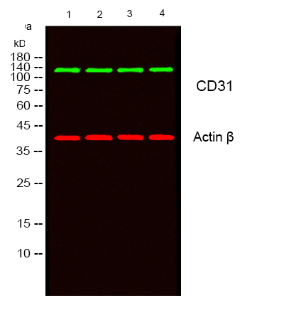

WB
Western blot analysis of lysates from 1) HepG2, 2) K562,3) L929,4) MOUSE-BRAIN cells, (Green) primary antibody was diluted at 1:1000, 4°over night, secondary antibody was diluted at 1:10000, 37° 1hour. (Red) Actin β Monoclonal Antibody antibody was diluted at 1:5000 as loading control, 4° over night,secondary antibody was diluted at 1:10000, 37° 1hour.IHC
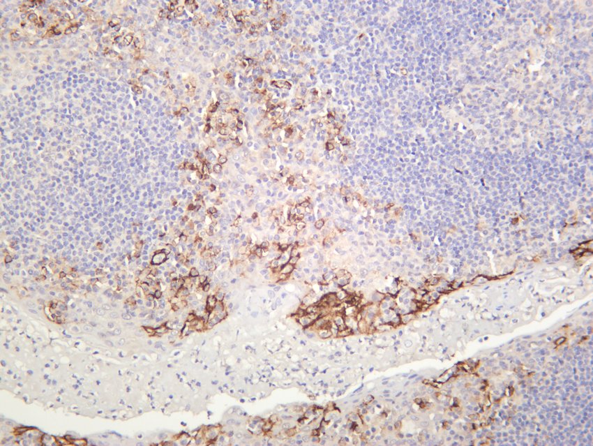
Human tonsil was stained with anti-CD31 rabbit antibody.IHC
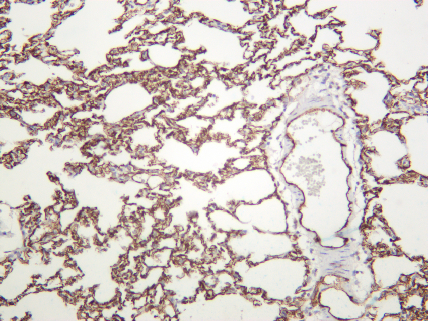
Mouse lung was stained with anti-CD31 rabbit antibody.IHC
Rat lung was stained with anti-CD31 rabbit antibody.ICC/IF
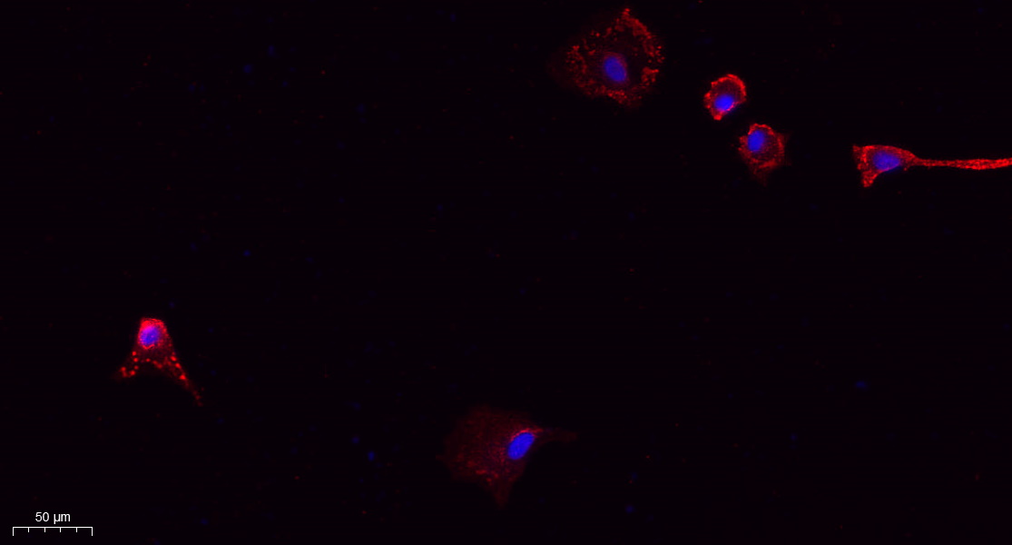
Immunofluorescence analysis of A549. 1,primary Antibody(red) was diluted at 1:200(4°C overnight). 2, Goat Anti Rabbit IgG (H&L) - Alexa Fluor 594 Secondary antibody was diluted at 1:1000(room temperature, 50min).3, Picture B: DAPI(blue) 10min.| Product Name | CD31 Rabbit mAb |
|---|---|
| Antibody Type | Primary Antibodies |
| Clonality | monoclonal |
|---|---|
| Isotype | IgG |
| Host Species | Rabbit |
| Tested Applications | ICC/IFIHCWB |
| WB:1:1000-1:5000 IHC:1:1000-1:2000 ICC/IF:1:200-1:1000 |
|
| Species Reactivity | HumanMouseRat |
| Concentration | 1mg/ml |
| Purification | Protein A |
| Gene Symbol | PECAM1 |
|---|---|
| Gene Synonyms | CD31 PECA1 GPIIA' PECAM-1 endoCAM CD31/EndoCAM |
| Gene Full Name | platelet and endothelial cell adhesion molecule 1 |
| Gene Summary | The protein encoded by this gene is found on the surface of platelets, monocytes, neutrophils, and some types of T-cells, and makes up a large portion of endothelial cell intercellular junctions. The encoded protein is a member of the immunoglobulin superfamily and is likely involved in leukocyte migration, angiogenesis, and integrin activation. [provided by RefSeq, May 2010] |
| Molecular Weight(MW) | 81kD (Calculated),130kD (Observed). |
| Cellular Localization | Membrane. |
WB
Western blot analysis of lysates from 1) HepG2, 2) K562,3) L929,4) MOUSE-BRAIN cells, (Green) primary antibody was diluted at 1:1000, 4°over night, secondary antibody was diluted at 1:10000, 37° 1hour. (Red) Actin β Monoclonal Antibody antibody was diluted at 1:5000 as loading control, 4° over night,secondary antibody was diluted at 1:10000, 37° 1hour.
IHC
Human tonsil was stained with anti-CD31 rabbit antibody.
IHC
Mouse lung was stained with anti-CD31 rabbit antibody.
IHC
Rat lung was stained with anti-CD31 rabbit antibody.
ICC/IF
Immunofluorescence analysis of A549. 1,primary Antibody(red) was diluted at 1:200(4°C overnight). 2, Goat Anti Rabbit IgG (H&L) - Alexa Fluor 594 Secondary antibody was diluted at 1:1000(room temperature, 50min).3, Picture B: DAPI(blue) 10min.| Application Notes | WB:1:1000-1:5000 IHC:1:1000-1:2000 ICC/IF:1:200-1:1000 |
|---|
| Form | Liquid |
|---|---|
| Storage Instructions | -15°C to -25°C/1 year(Do not lower than -25°C) |
| Storage Buffer | PBS, 50% glycerol, 0.05% Proclin 300, 0.05%BSA |


 Data sheet for OM644108