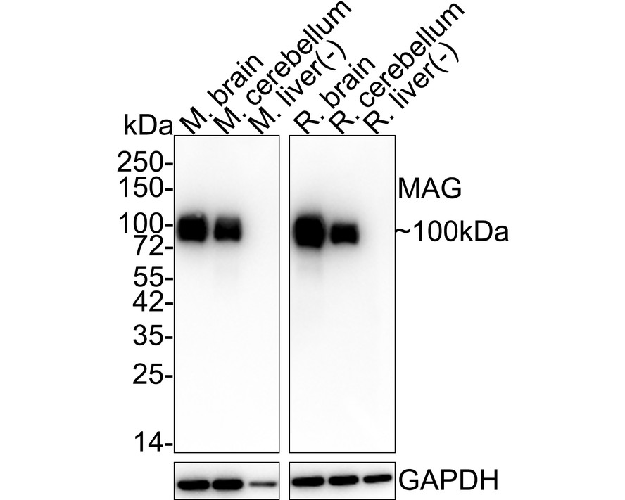
WB
Western blot analysis of MAG on different lysates with Rabbit anti-MAG antibody at 1/2,000 dilution. Lane 1: Mouse brain tissue lysate (no heat) (20 µg/Lane), Lane 2: Mouse cerebellum tissue lysate (20 µg/Lane), Lane 3: Mouse liver tissue lysate (negative) (20 µg/Lane), Lane 4: Rat brain tissue lysate (no heat) (20 µg/Lane), Lane 5: Rat cerebellum tissue lysate (20 µg/Lane), Lane 6: Rat liver tissue lysate (negative) (20 µg/Lane), Notice: no heat means the lysate is not boiled. Exposure time: 42 seconds; 4-20% SDS-PAGE gel. Proteins were transferred to a PVDF membrane and blocked with 5% NFDM/TBST for 1 hour at room temperature. The primary antibody at 1/2,000 dilution was used in 5% NFDM/TBST at room temperature for 2 hours. Goat Anti-Rabbit IgG - HRP Secondary Antibody at 1/50,000 dilution was used for 1 hour at room temperature.IHC
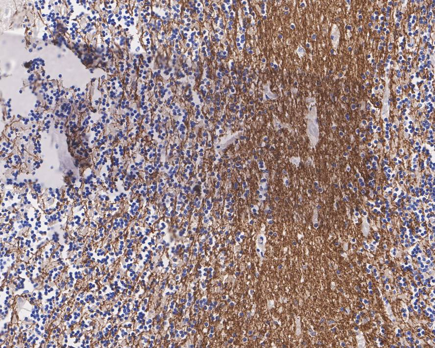
Immunohistochemical analysis of paraffin-embedded human cerebellum tissue with Rabbit anti-MAG antibody at 1/2,000 dilution. The section was pre-treated using heat mediated antigen retrieval with Tris-EDTA buffer (pH 9.0) for 20 minutes. The tissues were blocked in 1% BSA for 20 minutes at room temperature, washed with ddH2O and PBS, and then probed with the primary antibody at 1/2,000 dilution for 1 hour at room temperature. The detection was performed using an HRP conjugated compact polymer system. DAB was used as the chromogen. Tissues were counterstained with hematoxylin and mounted with DPX.IHC
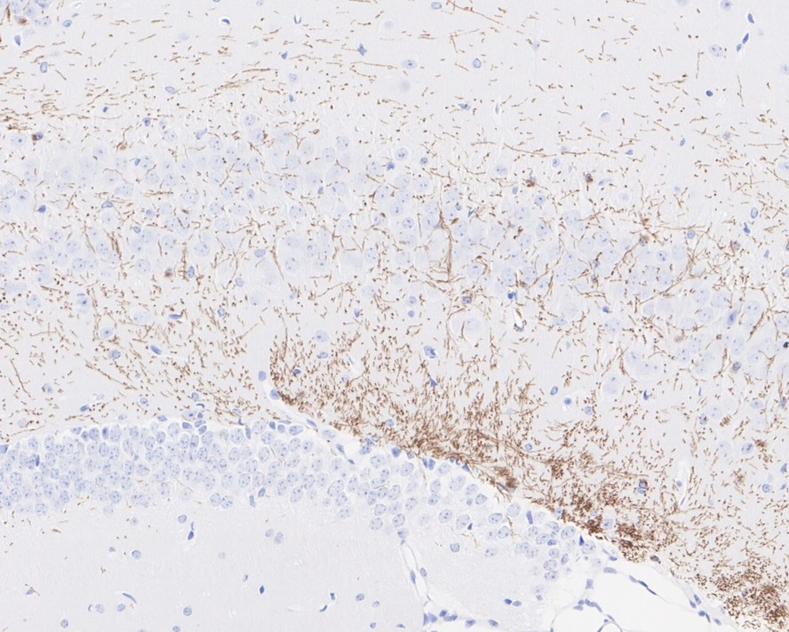
Immunohistochemical analysis of paraffin-embedded mouse hippocampus tissue with Rabbit anti-MAG antibody at 1/2,000 dilution. The section was pre-treated using heat mediated antigen retrieval with Tris-EDTA buffer (pH 9.0) for 20 minutes. The tissues were blocked in 1% BSA for 20 minutes at room temperature, washed with ddH2O and PBS, and then probed with the primary antibody at 1/2,000 dilution for 1 hour at room temperature. The detection was performed using an HRP conjugated compact polymer system. DAB was used as the chromogen. Tissues were counterstained with hematoxylin and mounted with DPX.IF-P
Species: Mouse, Site: Cerebellum, Sample: Paraffin-embedded section, Antibody concentration: 1:200.IF-F
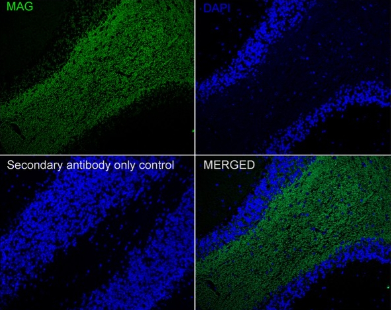
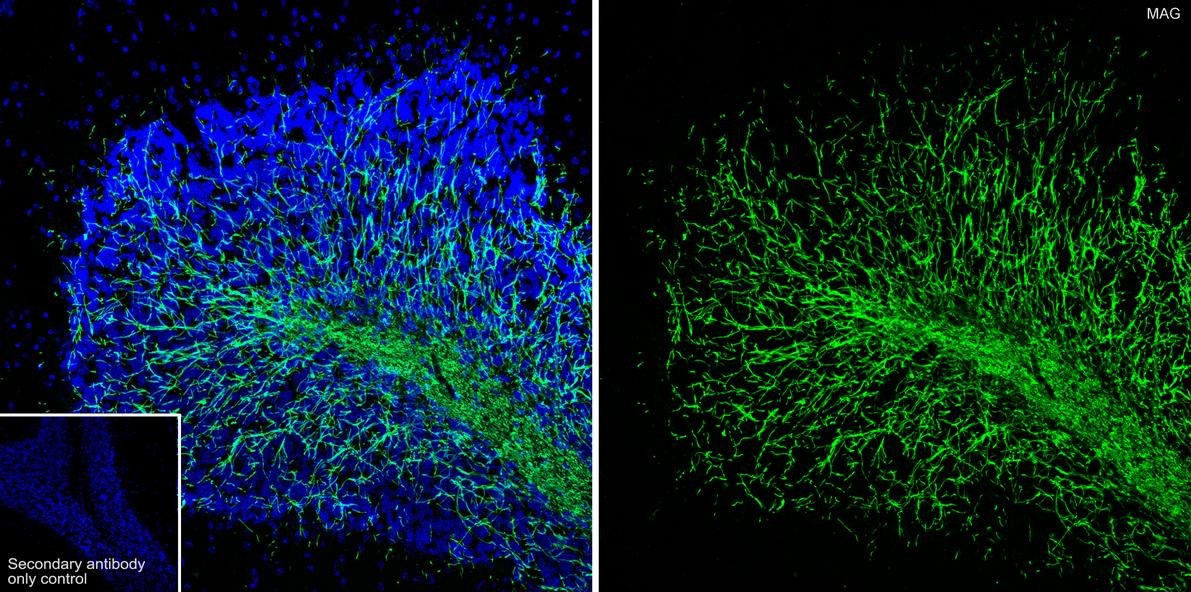
Species: Mouse, Site: Cerebellum, Sample: Frozen section, Antibody concentration: 1:500, Antigen retrieval: The section was pre-treated using heat mediated antigen retrieval with sodium citrate buffer (pH 6.0) for about 2 minutes in microwave oven.IP
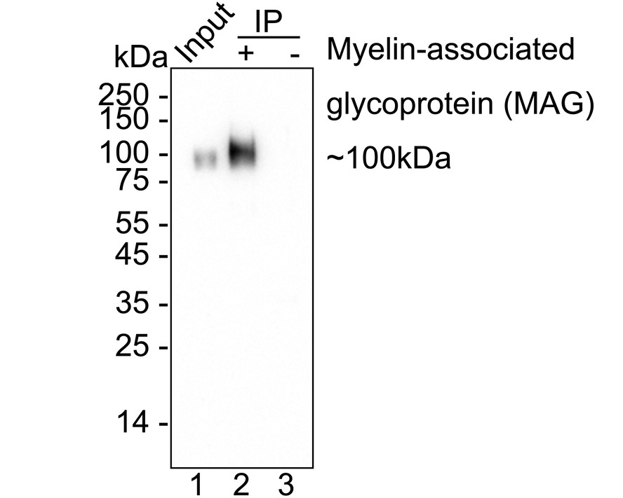
MAG was immunoprecipitated from 0.2 mg mouse brain tissue lysate with Rabbit anti-MAG antibody at 2 µg/25 µl agarose. Western blot was performed from the immunoprecipitate usingRabbit anti-MAG antibody at 1/1,000 dilution. Anti-Rabbit IgG for IP Nano-secondary antibody at 1/5,000 dilution was used for 1 hour at room temperature. Lane 1: Mouse brain tissue lysate (input), Lane 2: Rabbit anti-MAG antibody IP in mouse brain tissue lysate, Lane 3: Rabbit IgG instead of Rabbit anti-MAG antibody in mouse brain tissue lysate, Blocking/Dilution buffer: 5% NFDM/TBST Exposure time: 32 seconds.| Product Name | MAG Recombinant Rabbit Monoclonal Antibody |
|---|---|
| Antibody Type | Primary Antibodies |
| Immunogen | Recombinant protein within human MAG aa 1-536 / 626. |
| Clonality | monoclonal |
|---|---|
| Isotype | IgG |
| Host Species | Rabbit |
| Tested Applications | IF-FIF-PIHCIPWB |
| WB:1:2000 IHC:1:2000-1:5000 IF-P:1:200-1:1000 IF-F:1:500-1:1000 IP:1-2μg/sample |
|
| Species Reactivity | HumanMouseRat |
| Concentration | 1mg/ml |
| Purification | Protein A |
| Gene Symbol | MAG |
|---|---|
| Gene Synonyms | GMA S-MAG SPG75 SIGLEC4 SIGLEC4A SIGLEC-4A |
| Gene Full Name | myelin associated glycoprotein |
| Gene Summary | The protein encoded by this gene is a type I membrane protein and member of the immunoglobulin superfamily. It is thought to be involved in the process of myelination. It is a lectin that binds to sialylated glycoconjugates and mediates certain myelin-neuron cell-cell interactions. Three alternatively spliced transcripts encoding different isoforms have been described for this gene. [provided by RefSeq, Nov 2010] |
| Molecular Weight(MW) | 69kDa(Observed band size:100kDa) |
| Cellular Localization | Cell membrane, Membrane raft. |
WB
Western blot analysis of MAG on different lysates with Rabbit anti-MAG antibody at 1/2,000 dilution. Lane 1: Mouse brain tissue lysate (no heat) (20 µg/Lane), Lane 2: Mouse cerebellum tissue lysate (20 µg/Lane), Lane 3: Mouse liver tissue lysate (negative) (20 µg/Lane), Lane 4: Rat brain tissue lysate (no heat) (20 µg/Lane), Lane 5: Rat cerebellum tissue lysate (20 µg/Lane), Lane 6: Rat liver tissue lysate (negative) (20 µg/Lane), Notice: no heat means the lysate is not boiled. Exposure time: 42 seconds; 4-20% SDS-PAGE gel. Proteins were transferred to a PVDF membrane and blocked with 5% NFDM/TBST for 1 hour at room temperature. The primary antibody at 1/2,000 dilution was used in 5% NFDM/TBST at room temperature for 2 hours. Goat Anti-Rabbit IgG - HRP Secondary Antibody at 1/50,000 dilution was used for 1 hour at room temperature.
IHC
Immunohistochemical analysis of paraffin-embedded human cerebellum tissue with Rabbit anti-MAG antibody at 1/2,000 dilution. The section was pre-treated using heat mediated antigen retrieval with Tris-EDTA buffer (pH 9.0) for 20 minutes. The tissues were blocked in 1% BSA for 20 minutes at room temperature, washed with ddH2O and PBS, and then probed with the primary antibody at 1/2,000 dilution for 1 hour at room temperature. The detection was performed using an HRP conjugated compact polymer system. DAB was used as the chromogen. Tissues were counterstained with hematoxylin and mounted with DPX.
IHC
Immunohistochemical analysis of paraffin-embedded mouse hippocampus tissue with Rabbit anti-MAG antibody at 1/2,000 dilution. The section was pre-treated using heat mediated antigen retrieval with Tris-EDTA buffer (pH 9.0) for 20 minutes. The tissues were blocked in 1% BSA for 20 minutes at room temperature, washed with ddH2O and PBS, and then probed with the primary antibody at 1/2,000 dilution for 1 hour at room temperature. The detection was performed using an HRP conjugated compact polymer system. DAB was used as the chromogen. Tissues were counterstained with hematoxylin and mounted with DPX.
IF-P
Species: Mouse, Site: Cerebellum, Sample: Paraffin-embedded section, Antibody concentration: 1:200.
IF-F
Species: Mouse, Site: Cerebellum, Sample: Frozen section, Antibody concentration: 1:500, Antigen retrieval: The section was pre-treated using heat mediated antigen retrieval with sodium citrate buffer (pH 6.0) for about 2 minutes in microwave oven.
IP
MAG was immunoprecipitated from 0.2 mg mouse brain tissue lysate with Rabbit anti-MAG antibody at 2 µg/25 µl agarose. Western blot was performed from the immunoprecipitate usingRabbit anti-MAG antibody at 1/1,000 dilution. Anti-Rabbit IgG for IP Nano-secondary antibody at 1/5,000 dilution was used for 1 hour at room temperature. Lane 1: Mouse brain tissue lysate (input), Lane 2: Rabbit anti-MAG antibody IP in mouse brain tissue lysate, Lane 3: Rabbit IgG instead of Rabbit anti-MAG antibody in mouse brain tissue lysate, Blocking/Dilution buffer: 5% NFDM/TBST Exposure time: 32 seconds.| Application Notes | WB:1:2000 IHC:1:2000-1:5000 IF-P:1:200-1:1000 IF-F:1:500-1:1000 IP:1-2μg/sample |
|---|
| Form | Liquid |
|---|---|
| Storage Instructions | Store at +4℃ after thawing. Aliquot store at -20℃. Avoid repeated freeze / thaw cycles. |
| Storage Buffer | PBS (pH7.4), 0.1% BSA, 40% Glycerol. Preservative: 0.05% Sodium Azide. |


 Data sheet for OM644157