WB
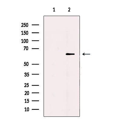
Western blot analysis of extracts from Human skin, using TRP1 Antibody. The lane on the left was treated with blocking peptide.IHC
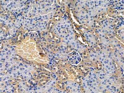
TRP1 Antibody at 1/100 staining mouse kidney tissue by IHC-P. The sample was formaldehyde fixed and a heat mediated antigen retrieval step in citrate buffer was performed. The sample was then blocked and incubated with the primary antibody at 4°C overnight. An HRP conjugated anti-rabbit antibody was used as the secondary antibody.| Product Name | Rabbit polyclonal antibody to TRP1 |
|---|---|
| Antibody Type | Primary Antibodies |
| Immunogen | A synthesized peptide derived from human TYRP1(Accession P17643), corresponding to amino acid residues F244-E294. |
| Clonality | Polyclonal |
|---|---|
| Isotype | IgG |
| Host Species | Rabbit |
| Tested Applications | IHCWB |
| WB:1:500-1:2000 IHC:1:50-1:200 |
|
| Species Reactivity | HumanMouseRat |
| Concentration | 1mg/ml |
| Purification | Affinity purified |
| Gene Symbol | TYRP1 |
|---|---|
| Gene Synonyms | TRP CAS2 CATB GP75 OCA3 TRP1 TYRP b-PROTEIN |
| Gene Full Name | tyrosinase related protein 1 |
| Gene Summary | This gene encodes a melanosomal enzyme that belongs to the tyrosinase family and plays an important role in the melanin biosynthetic pathway. Defects in this gene are the cause of rufous oculocutaneous albinism and oculocutaneous albinism type III. [provided by RefSeq, Mar 2009] |
| Alternative Names | 5 5 6 dihydroxyindole 2 carboxylic acid oxidase 6-dihydroxyindole-2-carboxylic acid oxidase b-PROTEIN CAS2 Catalase B CATB DHICA oxidase Glycoprotein 75 GP75 Melanoma antigen gp75 OCA3 TRP TRP-1 TRP1 Tyrosinase related protein 1 Tyrosinase-related protein 1 TYRP TYRP1 TYRP1_HUMAN TYRRP |
| Molecular Weight(MW) | 61kDa |
| Cellular Localization | Melanosome membrane>Single-pass type I membrane protein. Melanosome. |
WB
Western blot analysis of extracts from Human skin, using TRP1 Antibody. The lane on the left was treated with blocking peptide.
IHC
TRP1 Antibody at 1/100 staining mouse kidney tissue by IHC-P. The sample was formaldehyde fixed and a heat mediated antigen retrieval step in citrate buffer was performed. The sample was then blocked and incubated with the primary antibody at 4°C overnight. An HRP conjugated anti-rabbit antibody was used as the secondary antibody.| Application Notes | WB:1:500-1:2000 IHC:1:50-1:200 |
|---|
| Form | Liquid |
|---|---|
| Storage Instructions | Store at -20 °C. Stable for 12 months from date of receipt. |
| Storage Buffer | Rabbit IgG in phosphate buffered saline , pH 7.4, 150mM NaCl, 0.02% sodium azide and 50% glycerol. |


