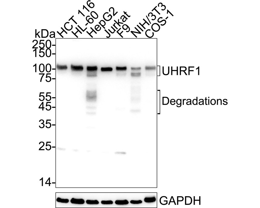
WB
Western blot analysis of UHRF1 on different lysates with Rabbit anti-UHRF1 antibody at 1/2,000 dilution. Lane 1: HCT 116 cell lysate, Lane 2: HL-60 cell lysate, Lane 3: HepG2 cell lysate, Lane 4: Jurkat cell lysate, Lane 5: F9 cell lysate, Lane 6: NIH/3T3 cell lysate, Lane 7: COS-1 cell lysate, Lysates/proteins at 20 µg/Lane. Exposure time: 10 seconds; 4-20% SDS-PAGE gel. Proteins were transferred to a PVDF membrane and blocked with 5% NFDM/TBST for 1 hour at room temperature. The primary antibody at 1/2,000 dilution was used in 5% NFDM/TBST at 4℃ overnight. Goat Anti-Rabbit IgG - HRP Secondary Antibody at 1/50,000 dilution was used for 1 hour at room temperature.ICC/IF
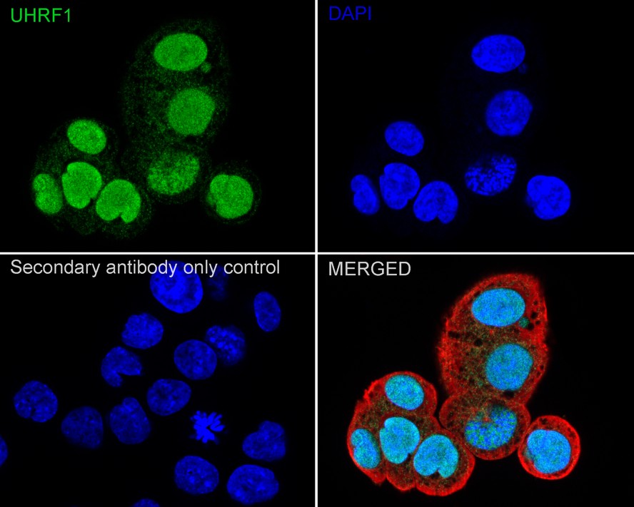
Immunocytochemistry analysis of HCT 116 cells labeling UHRF1 with Rabbit anti-UHRF1 antibody at 1/10,000 dilution. Cells were fixed in 4% paraformaldehyde for 15 minutes at room temperature, permeabilized with 0.1% Triton X-100 in PBS for 15 minutes at room temperature, then blocked with 1% BSA in 10% negative goat serum for 1 hour at room temperature. Cells were then incubated with Rabbit anti-UHRF1 antibody at 1/10,000 dilution in 1% BSA in PBST overnight at 4 ℃. Goat Anti-Rabbit IgG H&L (488) was used as the secondary antibody at 1/1,000 dilution. PBS instead of the primary antibody was used as the secondary antibody only control. Nuclear DNA was labelled in blue with DAPI. Beta tubulin (red) was stained at 1/100 dilution overnight at +4℃. Goat Anti-Mouse IgG H&L (594) was used as the secondary antibody at 1/1,000 dilution.FC
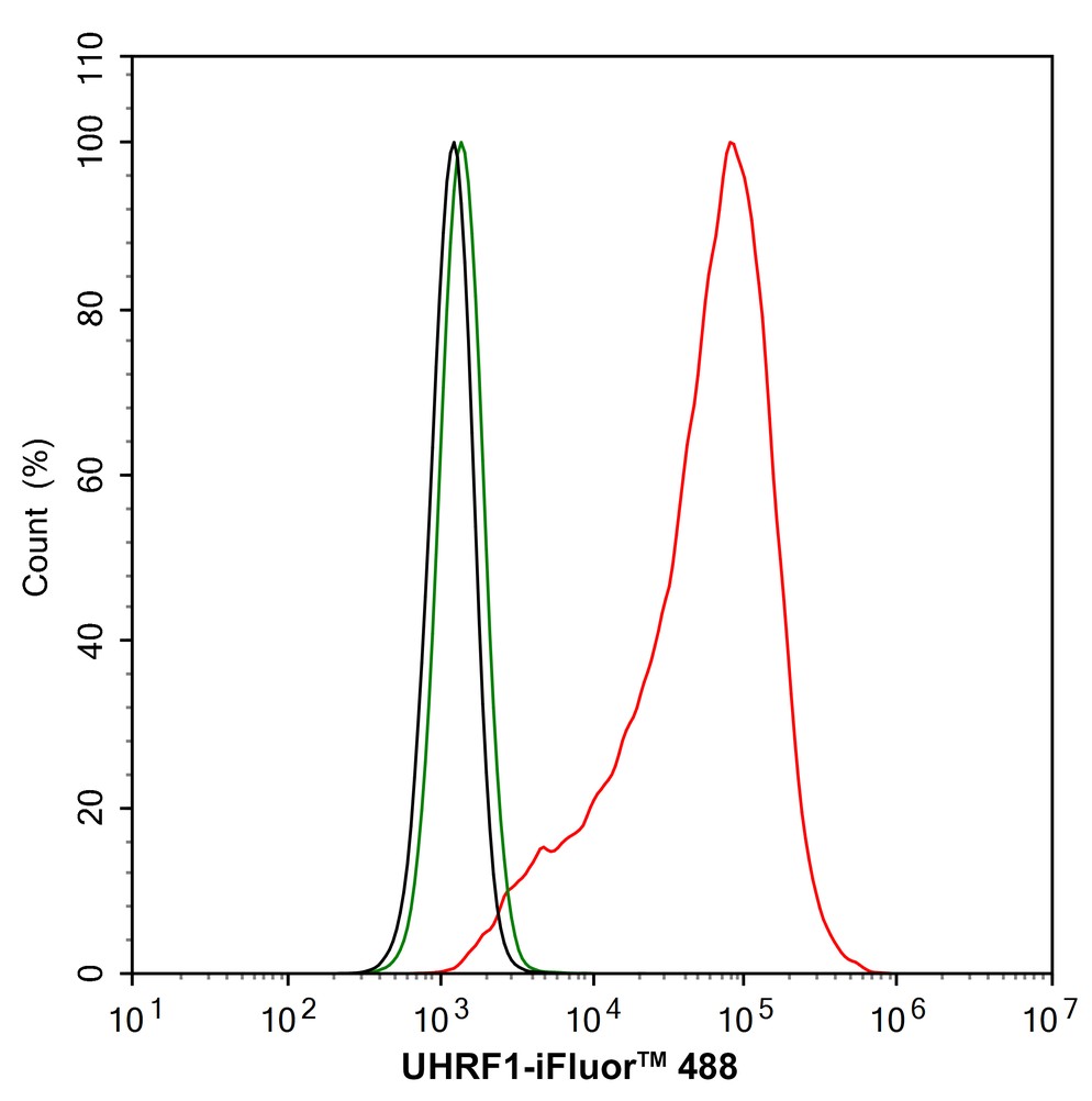
Flow cytometric analysis of HCT 116 cells labeling UHRF1. Cells were fixed and permeabilized. Then stained with the primary antibody (1/1,000) (red) compared with Rabbit IgG Isotype Control (green). After incubation of the primary antibody at +4℃ for an hour, the cells were stained with a iFluor™ 488 conjugate-Goat anti-Rabbit IgG Secondary antibody at 1/1,000 dilution for 30 minutes at +4℃. Unlabelled sample was used as a control (cells without incubation with primary antibody; black).IP
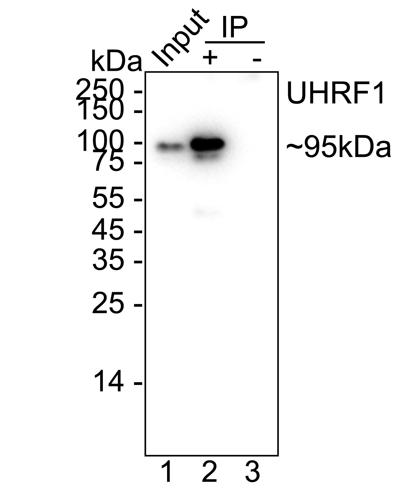
UHRF1 was immunoprecipitated from 0.2 mg HL-60 cell lysate with Rabbit anti-UHRF1 antibody at 2 µg/10 µl beads. Western blot was performed from the immunoprecipitate using Rabbit anti-UHRF1 antibody at 1/1,000 dilution. HRP Conjugated Anti-Rabbit IgG for IP Nano-secondary antibody at 1/5,000 dilution was used for 1 hour at room temperature. Lane 1: HL-60 cell lysate (input), Lane 2:Rabbit anti-UHRF1 antibody IP in HL-60 cell lysate, Lane 3: Rabbit IgG instead of Rabbit anti-UHRF1 antibody in HL-60 cell lysate, Blocking/Dilution buffer: 5% NFDM/TBST, Exposure time: 9 seconds.| Product Name | UHRF1 Recombinant Rabbit Monoclonal Antibody |
|---|---|
| Antibody Type | Primary Antibodies |
| Immunogen | Recombinant protein within human UHRF1 aa 1-350. |
| Clonality | monoclonal |
|---|---|
| Isotype | IgG |
| Host Species | Rabbit |
| Tested Applications | FCICC/IFIPWB |
| WB:1:2000 ICC/IF:1:10000 FC:1:1000 IP:1-2μg/sample |
|
| Species Reactivity | HumanMonkeyMouse |
| Concentration | 1mg/ml |
| Purification | Protein A |
| Gene Symbol | UHRF1 |
|---|---|
| Gene Synonyms | Np95 hNP95 ICBP90 RNF106 TDRD22 hUHRF1 huNp95 |
| Gene Full Name | ubiquitin like with PHD and ring finger domains 1 |
| Gene Summary | This gene encodes a member of a subfamily of RING-finger type E3 ubiquitin ligases. The protein binds to specific DNA sequences, and recruits a histone deacetylase to regulate gene expression. Its expression peaks at late G1 phase and continues during G2 and M phases of the cell cycle. It plays a major role in the G1/S transition by regulating topoisomerase IIalpha and retinoblastoma gene expression, and functions in the p53-dependent DNA damage checkpoint. It is regarded as a hub protein for the integration of epigenetic information. This gene is up-regulated in various cancers, and it is therefore considered to be a therapeutic target. Multiple transcript variants encoding different isoforms have been found for this gene. A related pseudogene exists on chromosome 12. [provided by RefSeq, Feb 2014] |
| Molecular Weight(MW) | 90kDa(Observed band size: 90-100kDa) |
| Cellular Localization | Nucleus. |
WB
Western blot analysis of UHRF1 on different lysates with Rabbit anti-UHRF1 antibody at 1/2,000 dilution. Lane 1: HCT 116 cell lysate, Lane 2: HL-60 cell lysate, Lane 3: HepG2 cell lysate, Lane 4: Jurkat cell lysate, Lane 5: F9 cell lysate, Lane 6: NIH/3T3 cell lysate, Lane 7: COS-1 cell lysate, Lysates/proteins at 20 µg/Lane. Exposure time: 10 seconds; 4-20% SDS-PAGE gel. Proteins were transferred to a PVDF membrane and blocked with 5% NFDM/TBST for 1 hour at room temperature. The primary antibody at 1/2,000 dilution was used in 5% NFDM/TBST at 4℃ overnight. Goat Anti-Rabbit IgG - HRP Secondary Antibody at 1/50,000 dilution was used for 1 hour at room temperature.
ICC/IF
Immunocytochemistry analysis of HCT 116 cells labeling UHRF1 with Rabbit anti-UHRF1 antibody at 1/10,000 dilution. Cells were fixed in 4% paraformaldehyde for 15 minutes at room temperature, permeabilized with 0.1% Triton X-100 in PBS for 15 minutes at room temperature, then blocked with 1% BSA in 10% negative goat serum for 1 hour at room temperature. Cells were then incubated with Rabbit anti-UHRF1 antibody at 1/10,000 dilution in 1% BSA in PBST overnight at 4 ℃. Goat Anti-Rabbit IgG H&L (488) was used as the secondary antibody at 1/1,000 dilution. PBS instead of the primary antibody was used as the secondary antibody only control. Nuclear DNA was labelled in blue with DAPI. Beta tubulin (red) was stained at 1/100 dilution overnight at +4℃. Goat Anti-Mouse IgG H&L (594) was used as the secondary antibody at 1/1,000 dilution.
FC
Flow cytometric analysis of HCT 116 cells labeling UHRF1. Cells were fixed and permeabilized. Then stained with the primary antibody (1/1,000) (red) compared with Rabbit IgG Isotype Control (green). After incubation of the primary antibody at +4℃ for an hour, the cells were stained with a iFluor™ 488 conjugate-Goat anti-Rabbit IgG Secondary antibody at 1/1,000 dilution for 30 minutes at +4℃. Unlabelled sample was used as a control (cells without incubation with primary antibody; black).
IP
UHRF1 was immunoprecipitated from 0.2 mg HL-60 cell lysate with Rabbit anti-UHRF1 antibody at 2 µg/10 µl beads. Western blot was performed from the immunoprecipitate using Rabbit anti-UHRF1 antibody at 1/1,000 dilution. HRP Conjugated Anti-Rabbit IgG for IP Nano-secondary antibody at 1/5,000 dilution was used for 1 hour at room temperature. Lane 1: HL-60 cell lysate (input), Lane 2:Rabbit anti-UHRF1 antibody IP in HL-60 cell lysate, Lane 3: Rabbit IgG instead of Rabbit anti-UHRF1 antibody in HL-60 cell lysate, Blocking/Dilution buffer: 5% NFDM/TBST, Exposure time: 9 seconds.| Application Notes | WB:1:2000 ICC/IF:1:10000 FC:1:1000 IP:1-2μg/sample |
|---|
| Form | Liquid |
|---|---|
| Storage Instructions | Store at +4℃ after thawing. Aliquot store at -20℃. Avoid repeated freeze / thaw cycles. |
| Storage Buffer | PBS (pH7.4), 0.1% BSA, 40% Glycerol. Preservative: 0.05% Sodium Azide. |


 Data sheet for OM644269