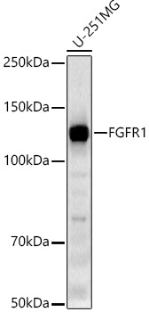
WB
Western blot analysis of lysates from U-251MG cells, using FGFR1 Rabbit mAb at 1:10000 dilution. Secondary antibody: HRP-conjugated Goat anti-Rabbit IgG (H+L) at 1:10000 dilution. Lysates/proteins: 25μg per lane. Blocking buffer: 3% nonfat dry milk in TBST. Detection: ECL Basic Kit. Exposure time: 180s.WB
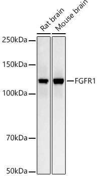
Western blot analysis of various lysates, using FGFR1 Rabbit mAb at 1:10000 dilution. Secondary antibody: HRP-conjugated Goat anti-Rabbit IgG (H+L)at 1:10000 dilution. Lysates/proteins: 25μg per lane. Blocking buffer: 3% nonfat dry milk in TBST. Detection: ECL Basic Kit. Exposure time: 10s.IHC
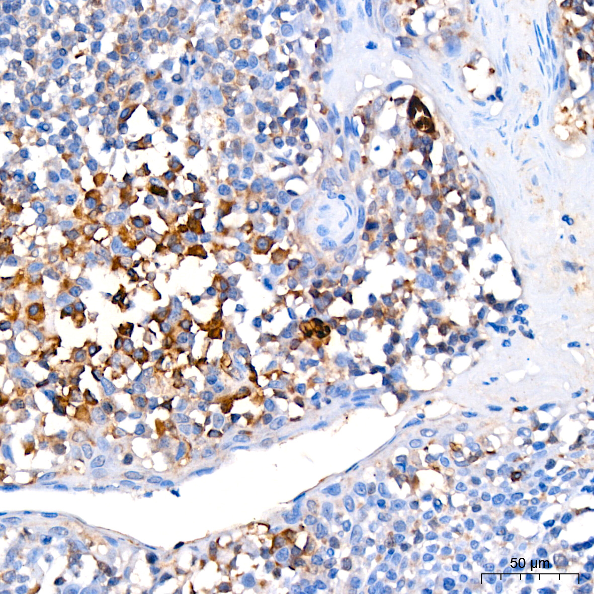
Immunohistochemistry analysis of paraffin embedded Human tonsil using FGFR1 Rabbit mAb at dilution of 1:200 (40x lens). High pressure antigen retrieval performed with 0.01M Citrate Bufferr (pH 6.0) prior to IHC staining.IHC
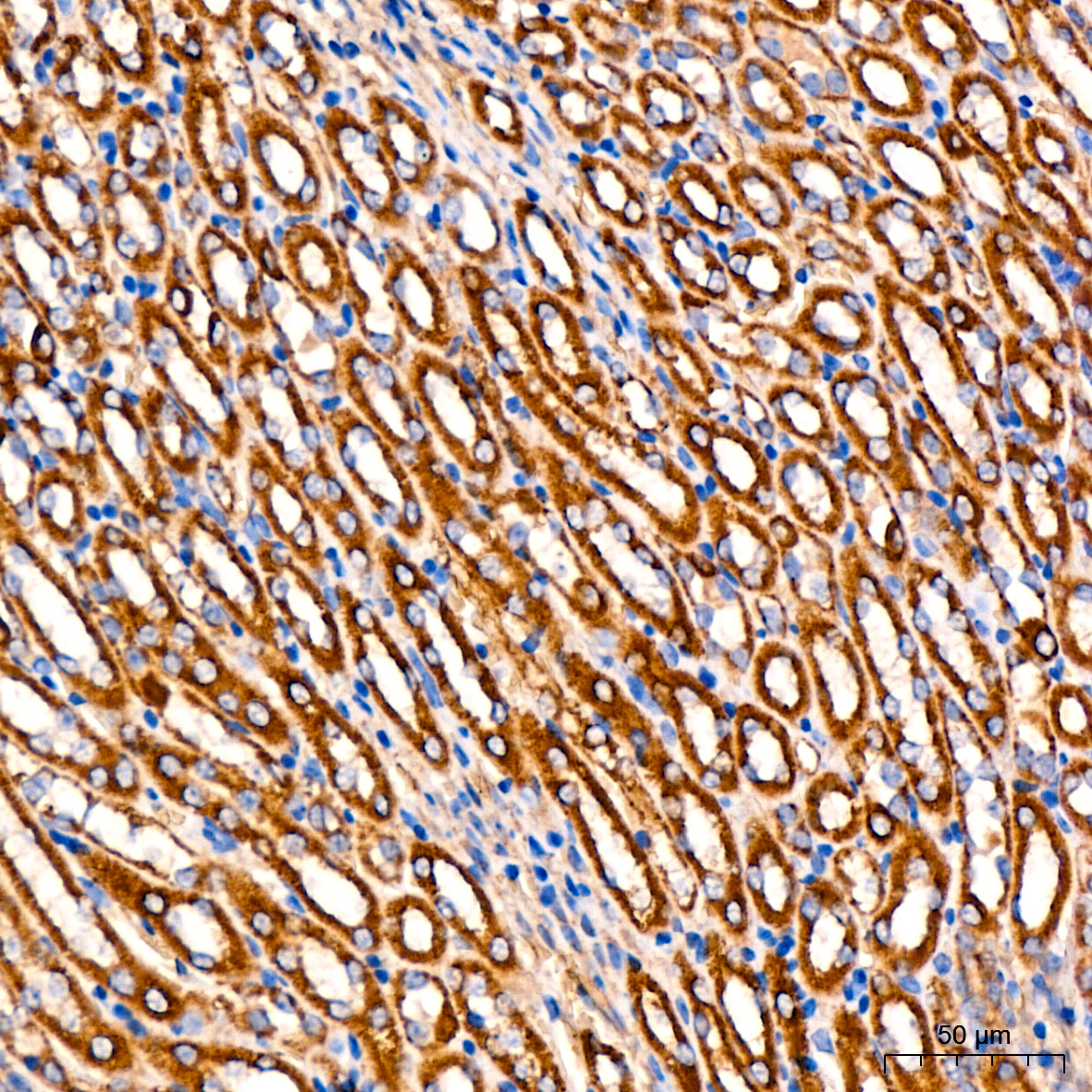
Immunohistochemistry analysis of paraffin embedded Mouse kidney using FGFR1 Rabbit mAb at dilution of 1:200 (40x lens). High pressure antigen retrieval performed with 0.01M Citrate Bufferr (pH 6.0) prior to IHC staining.ICC/IF
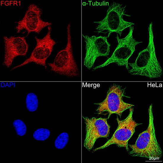
Confocal imaging of HeLa cells using FGFR1 Rabbit mAb (dilution 1:200)(Red). The cells were counterstained with α-Tubulin Mouse mAb (dilution 1:400) (Green). DAPI was used for nuclear staining (blue). Objective: 100x.| Product Name | FGFR1 Rabbit mAb |
|---|---|
| Antibody Type | Primary Antibodies |
| Immunogen | A synthetic peptide corresponding to a sequence within amino acids 600-700 of human FGFR1 (NP_075598.2). |
| Clonality | monoclonal |
|---|---|
| Isotype | IgG |
| Host Species | Rabbit |
| Tested Applications | ICC/IFIHCWB |
| WB:1:5000-1:10000 IHC:1:200-1:800 ICC/IF:1:50-1:200 |
|
| Species Reactivity | HumanMouseRat |
| Concentration | 1mg/ml |
| Purification | Affinity purified |
| Gene Symbol | FGFR1 |
|---|---|
| Gene Synonyms | CEK FLG HH2 OGD ECCL FLT2 KAL2 BFGFR CD331 FGFBR FLT-2 HBGFR N-SAM FGFR-1 HRTFDS bFGF-R-1 |
| Gene Full Name | fibroblast growth factor receptor 1 |
| Gene Summary | The protein encoded by this gene is a member of the fibroblast growth factor receptor (FGFR) family, where amino acid sequence is highly conserved between members and throughout evolution. FGFR family members differ from one another in their ligand affinities and tissue distribution. A full-length representative protein consists of an extracellular region, composed of three immunoglobulin-like domains, a single hydrophobic membrane-spanning segment and a cytoplasmic tyrosine kinase domain. The extracellular portion of the protein interacts with fibroblast growth factors, setting in motion a cascade of downstream signals, ultimately influencing mitogenesis and differentiation. This particular family member binds both acidic and basic fibroblast growth factors and is involved in limb induction. Mutations in this gene have been associated with Pfeiffer syndrome, Jackson-Weiss syndrome, Antley-Bixler syndrome, osteoglophonic dysplasia, and autosomal dominant Kallmann syndrome 2. Chromosomal aberrations involving this gene are associated with stem cell myeloproliferative disorder and stem cell leukemia lymphoma syndrome. Alternatively spliced variants which encode different protein isoforms have been described; however, not all variants have been fully characterized. [provided by RefSeq, Jul 2008] |
| Molecular Weight(MW) | 92kDa(Observed MW 122kDa) |
| Cellular Localization | Cell membrane, Cytoplasm, Cytoplasmic vesicle, Nucleus, Single-pass type I membrane protein, cytosol. |
WB
Western blot analysis of lysates from U-251MG cells, using FGFR1 Rabbit mAb at 1:10000 dilution. Secondary antibody: HRP-conjugated Goat anti-Rabbit IgG (H+L) at 1:10000 dilution. Lysates/proteins: 25μg per lane. Blocking buffer: 3% nonfat dry milk in TBST. Detection: ECL Basic Kit. Exposure time: 180s.
WB
Western blot analysis of various lysates, using FGFR1 Rabbit mAb at 1:10000 dilution. Secondary antibody: HRP-conjugated Goat anti-Rabbit IgG (H+L)at 1:10000 dilution. Lysates/proteins: 25μg per lane. Blocking buffer: 3% nonfat dry milk in TBST. Detection: ECL Basic Kit. Exposure time: 10s.
IHC
Immunohistochemistry analysis of paraffin embedded Human tonsil using FGFR1 Rabbit mAb at dilution of 1:200 (40x lens). High pressure antigen retrieval performed with 0.01M Citrate Bufferr (pH 6.0) prior to IHC staining.
IHC
Immunohistochemistry analysis of paraffin embedded Mouse kidney using FGFR1 Rabbit mAb at dilution of 1:200 (40x lens). High pressure antigen retrieval performed with 0.01M Citrate Bufferr (pH 6.0) prior to IHC staining.
ICC/IF
Confocal imaging of HeLa cells using FGFR1 Rabbit mAb (dilution 1:200)(Red). The cells were counterstained with α-Tubulin Mouse mAb (dilution 1:400) (Green). DAPI was used for nuclear staining (blue). Objective: 100x.| Application Notes | WB:1:5000-1:10000 IHC:1:200-1:800 ICC/IF:1:50-1:200 |
|---|
| Form | Liquid |
|---|---|
| Storage Instructions | Store at -20℃. Avoid freeze / thaw cycles. |
| Storage Buffer | Buffer: PBS with 0.05% proclin300, 0.05% BSA, 50% glycerol, pH7.3. |


 Data sheet for OM644279