WB
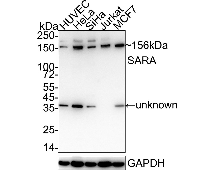
Western blot analysis of SARA on different lysates with Rabbit anti-SARA antibody at 1/1,000 dilution. Lane 1: HUVEC cell lysate, Lane 2: HeLa cell lysate, Lane 3: SiHa cell lysate, Lane 4: Jurkat cell lysate, Lane 5: MCF7 cell lysate, Lysates/proteins at 20 µg/Lane. Exposure time: 3 minutes; 4-20% SDS-PAGE gel. Proteins were transferred to a PVDF membrane and blocked with 5% NFDM/TBST for 1 hour at room temperature. The primary antibody at 1/1,000 dilution was used in 5% NFDM/TBST at 4℃ overnight. Goat Anti-Rabbit IgG - HRP Secondary Antibody at 1/50,000 dilution was used for 1 hour at room temperature.WB
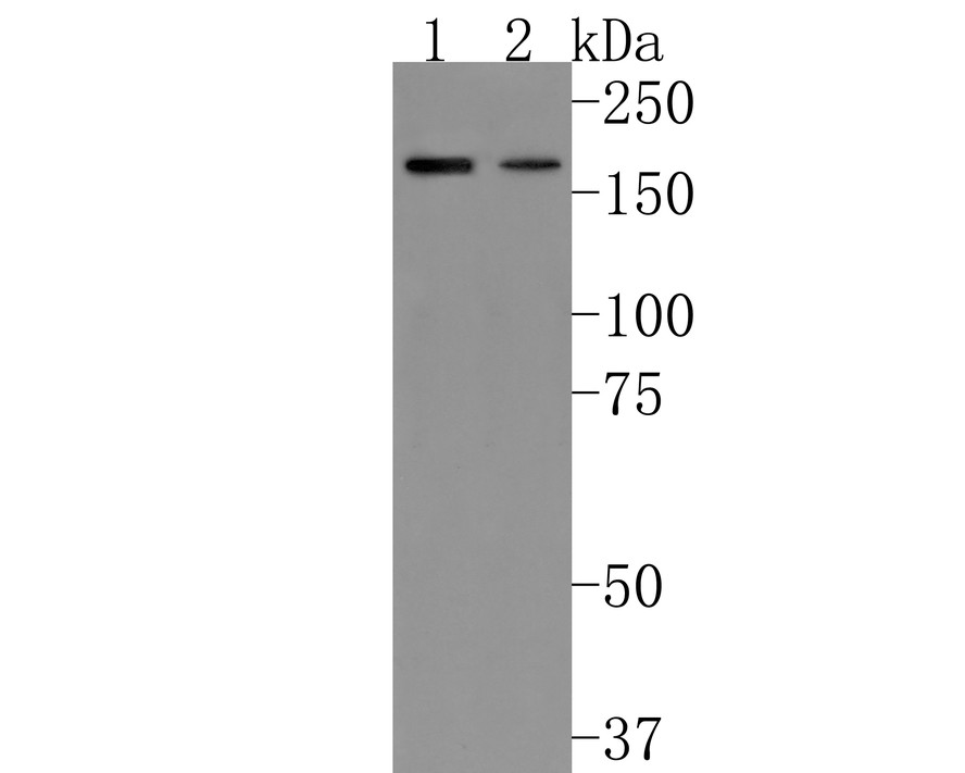
Western blot analysis of SARA on different lysates with Rabbit anti-SARA antibody at 1/500 dilution. Lane 1: HUVEC cell lysate, 10 µg/Lane, Lane 2: Rat brain tissue lysate, 20 µg/Lane, Exposure time: 2 minutes; 8% SDS-PAGE gel. Proteins were transferred to a PVDF membrane and blocked with 5% NFDM/TBST for 1 hour at room temperature. The primary antibody at 1/500 dilution was used in 5% NFDM/TBST at room temperature for 2 hours. Goat Anti-Rabbit IgG - HRP Secondary Antibody at 1:200,000 dilution was used for 1 hour at room temperature.| Product Name | SARA Recombinant Rabbit Monoclonal Antibody |
|---|---|
| Antibody Type | Primary Antibodies |
| Immunogen | Synthetic peptide within human SARA aa 1-50/1,425. |
| Clonality | monoclonal |
|---|---|
| Isotype | IgG |
| Host Species | Rabbit |
| Tested Applications | WB |
| WB:1:1000 |
|
| Species Reactivity | HumanRat |
| Concentration | 1mg/ml |
| Purification | Protein A |
| Gene Symbol | ZFYVE9 |
|---|---|
| Gene Synonyms | NSP SARA MADHIP SMADIP PPP1R173 |
| Gene Full Name | zinc finger FYVE-type containing 9 |
| Gene Summary | This gene encodes a double zinc finger motif-containing protein that participates in the transforming growth factor-beta (TGFB) signalling pathway. The encoded protein interacts directly with SMAD2 and SMAD3, and recruits SMAD2 to the TGFB receptor. There are multiple pseudogenes for this gene on chromosomes 2, 15, and X. Alternative splicing results in multiple transcript variants. [provided by RefSeq, Mar 2013] |
| Molecular Weight(MW) | 156kDa |
| Cellular Localization | Early endosome membrane, Cytoplasm. |
WB
Western blot analysis of SARA on different lysates with Rabbit anti-SARA antibody at 1/1,000 dilution. Lane 1: HUVEC cell lysate, Lane 2: HeLa cell lysate, Lane 3: SiHa cell lysate, Lane 4: Jurkat cell lysate, Lane 5: MCF7 cell lysate, Lysates/proteins at 20 µg/Lane. Exposure time: 3 minutes; 4-20% SDS-PAGE gel. Proteins were transferred to a PVDF membrane and blocked with 5% NFDM/TBST for 1 hour at room temperature. The primary antibody at 1/1,000 dilution was used in 5% NFDM/TBST at 4℃ overnight. Goat Anti-Rabbit IgG - HRP Secondary Antibody at 1/50,000 dilution was used for 1 hour at room temperature.
WB
Western blot analysis of SARA on different lysates with Rabbit anti-SARA antibody at 1/500 dilution. Lane 1: HUVEC cell lysate, 10 µg/Lane, Lane 2: Rat brain tissue lysate, 20 µg/Lane, Exposure time: 2 minutes; 8% SDS-PAGE gel. Proteins were transferred to a PVDF membrane and blocked with 5% NFDM/TBST for 1 hour at room temperature. The primary antibody at 1/500 dilution was used in 5% NFDM/TBST at room temperature for 2 hours. Goat Anti-Rabbit IgG - HRP Secondary Antibody at 1:200,000 dilution was used for 1 hour at room temperature.| Application Notes | WB:1:1000 |
|---|
| Form | Liquid |
|---|---|
| Storage Instructions | Store at +4℃ after thawing. Aliquot store at -20℃. Avoid repeated freeze / thaw cycles. |
| Storage Buffer | 1*TBS (pH7.4), 0.05% BSA, 40% Glycerol. Preservative: 0.05% Sodium Azide. |


 Data sheet for OM644307