WB
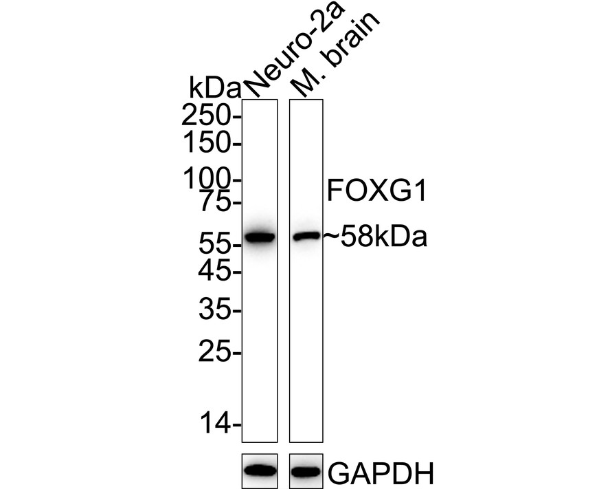
Western blot analysis of FOXG1 on different lysates with Rabbit anti-FOXG1 antibody at 1/2,000 dilution. Lane 1: Neuro-2a cell lysate (20 µg/Lane), Lane 2: Mouse brain tissue lysate (40 µg/Lane), Exposure time: Lane 1: 10 seconds; Lane 2: 3 minutes; 4-20% SDS-PAGE gel. Proteins were transferred to a PVDF membrane and blocked with 5% NFDM/TBST for 1 hour at room temperature. The primary antibody at 1/2,000 dilution was used in 5% NFDM/TBST at 4℃ overnight. Goat Anti-Rabbit IgG - HRP Secondary Antibody at 1/50,000 dilution was used for 1 hour at room temperature.IP
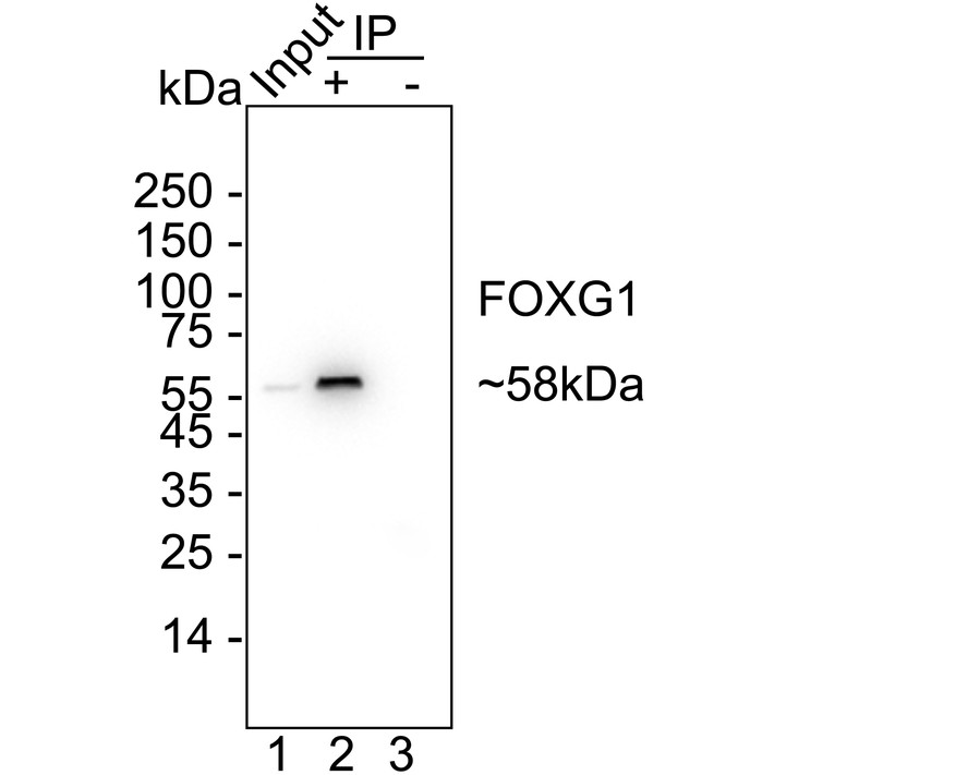
FOXG1 was immunoprecipitated from 0.2 mg Neuro-2a cell lysate with Rabbit anti-FOXG1 antibody at 2 µg/25 µl agarose. Western blot was performed from the immunoprecipitate using Rabbit anti-FOXG1 antibody at 1/2,000 dilution. Anti-Rabbit IgG for IP Nano-secondary antibody at 1/5,000 dilution was used for 1 hour at room temperature. Lane 1: Neuro-2a cell lysate (input), Lane 2:Rabbit anti-FOXG1 antibody IP in Neuro-2a cell lysate, Lane 3: Rabbit IgG instead of Rabbit anti-FOXG1 antibody in Neuro-2a cell lysate, Blocking/Dilution buffer: 5% NFDM/TBST, Exposure time: 3 seconds.| Product Name | FOXG1 Recombinant Rabbit Monoclonal Antibody |
|---|---|
| Antibody Type | Primary Antibodies |
| Immunogen | Recombinant protein within mouse FOXG1 aa 1-200. |
| Clonality | monoclonal |
|---|---|
| Isotype | IgG |
| Host Species | Rabbit |
| Tested Applications | IPWB |
| WB:1:2000 IP:1-2μg/sample |
|
| Species Reactivity | Mouse |
| Concentration | 1mg/ml |
| Purification | Protein A |
| Gene Symbol | Foxg1 |
|---|---|
| Gene Synonyms | Bf1 BF-1 Hfh9 Hfhbf1 2900064B05Rik |
| Gene Full Name | forkhead box G1 |
| Gene Summary | Enables sequence-specific DNA binding activity. Acts upstream of or within several processes, including neuron differentiation; positive regulation of neuroblast proliferation; and regulation of neuron differentiation. Located in nucleus. Is expressed in several structures, including central nervous system; early embryo; embryo ectoderm; gonad; and sensory organ. Used to study Rett syndrome. Orthologous to human FOXG1 (forkhead box G1). [provided by Alliance of Genome Resources, May 2025] |
| Alternative Names | BF-1 antibody BF-2 antibody BF1 antibody BF2 antibody Brain factor 1 antibody Brain factor 2 antibody FHKL antibody FKH2 antibody FKHL1 antibody FKHL2 antibody BF-1 antibody BF-2 antibody BF1 antibody BF2 antibody Brain factor 1 antibody Brain factor 2 antibody FHKL antibody FKH2 antibody FKHL1 antibody FKHL2 antibody FKHL3 antibody FKHL4 antibody Forkhead box protein G1 antibody Forkhead box protein G1A antibody Forkhead box protein G1B antibody Forkhead box protein G1C antibody Forkhead like 1 antibody Forkhead like 2 antibody Forkhead like 3 antibody Forkhead like 4 antibody Forkhead-related protein FKHL1 antibody Forkhead-related protein FKHL2 antibody Forkhead-related protein FKHL3 antibody FOXG1 antibody FOXG1_HUMAN antibody FOXG1A antibody FOXG1B antibody FOXG1C antibody HBF 1 antibody HBF G2 antibody hBF-2 antibody HBF2 antibody HFK1 antibody HFK2 antibody HFK3 antibody KHL2 antibody Oncogene QIN antibody QIN antibody |
| Molecular Weight(MW) | 52kDa(Observed band size: 58kDa) |
| Cellular Localization | Nucleus. |
WB
Western blot analysis of FOXG1 on different lysates with Rabbit anti-FOXG1 antibody at 1/2,000 dilution. Lane 1: Neuro-2a cell lysate (20 µg/Lane), Lane 2: Mouse brain tissue lysate (40 µg/Lane), Exposure time: Lane 1: 10 seconds; Lane 2: 3 minutes; 4-20% SDS-PAGE gel. Proteins were transferred to a PVDF membrane and blocked with 5% NFDM/TBST for 1 hour at room temperature. The primary antibody at 1/2,000 dilution was used in 5% NFDM/TBST at 4℃ overnight. Goat Anti-Rabbit IgG - HRP Secondary Antibody at 1/50,000 dilution was used for 1 hour at room temperature.
IP
FOXG1 was immunoprecipitated from 0.2 mg Neuro-2a cell lysate with Rabbit anti-FOXG1 antibody at 2 µg/25 µl agarose. Western blot was performed from the immunoprecipitate using Rabbit anti-FOXG1 antibody at 1/2,000 dilution. Anti-Rabbit IgG for IP Nano-secondary antibody at 1/5,000 dilution was used for 1 hour at room temperature. Lane 1: Neuro-2a cell lysate (input), Lane 2:Rabbit anti-FOXG1 antibody IP in Neuro-2a cell lysate, Lane 3: Rabbit IgG instead of Rabbit anti-FOXG1 antibody in Neuro-2a cell lysate, Blocking/Dilution buffer: 5% NFDM/TBST, Exposure time: 3 seconds.| Application Notes | WB:1:2000 IP:1-2μg/sample |
|---|
| Form | Liquid |
|---|---|
| Storage Instructions | Store at +4℃ after thawing. Aliquot store at -20℃. Avoid repeated freeze / thaw cycles. |
| Storage Buffer | PBS (pH7.4), 0.1% BSA, 40% Glycerol. Preservative: 0.05% Sodium Azide. |


