WB
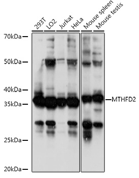
Western blot analysis of various lysates using MTHFD2 Rabbit pAb at 1:1000 dilution. Secondary antibody: HRP-conjugated Goat anti-Rabbit IgG (H+L) at 1:10000 dilution. Lysates/proteins: 25μg per lane. Blocking buffer: 3% nonfat dry milk in TBST. Detection: ECL Basic Kit. Exposure time: 30s.IHC
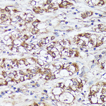
Immunohistochemistry analysis of paraffin embedded Human liver cancer using MTHFD2 Rabbit pAb at dilution of 1:100 (40x lens). Microwave antigen retrieval performed with 0.01M PBS Buffer (pH 7.2) prior to IHC staining.IHC
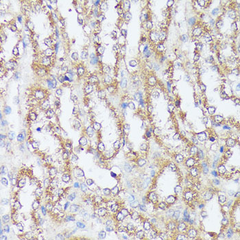
Immunohistochemistry analysis of paraffin embedded Rat kidney using MTHFD2 Rabbit pAb at dilution of 1:100 (40x lens). Microwave antigen retrieval performed with 0.01M PBS Buffer (pH 7.2) prior to IHC staining.IHC
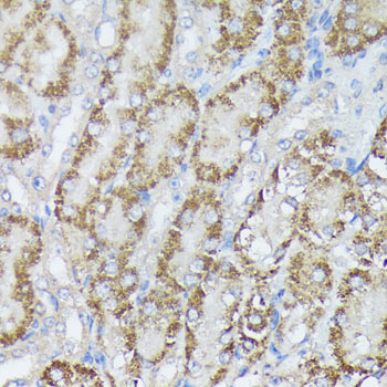
Immunohistochemistry analysis of paraffin embedded Mouse kidney using MTHFD2 Rabbit pAb at dilution of 1:100 (40x lens). Microwave antigen retrieval performed with 0.01M PBS Buffer (pH 7.2) prior to IHC staining.ICC/IF
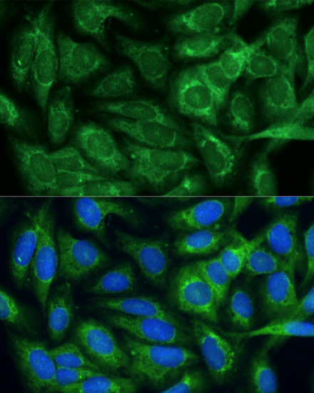
Immunofluorescence analysis of U-2 OS cells using MTHFD2 Rabbit pAb at dilution of 1:100. Secondary antibody: Omnimabs® 488 Goat Anti-Rabbit IgG(H&L) at 1:500 dilution. Blue: DAPI for nuclear staining.| Product Name | MTHFD2 Rabbit pAb |
|---|---|
| Antibody Type | Primary Antibodies |
| Immunogen | Recombinant fusion protein containing a sequence corresponding to amino acids 40-250 of human MTHFD2 (NP_006627.2). |
| Clonality | Polyclonal |
|---|---|
| Isotype | IgG |
| Host Species | Rabbit |
| Tested Applications | ICC/IFIHCWB |
| WB:1:500-1:1000 IHC:1:50-1:200 ICC/IF:1:50-1:200 |
|
| Species Reactivity | HumanMouseRat |
| Concentration | 1mg/ml |
| Purification | Affinity purified |
| Gene Symbol | MTHFD2 |
|---|---|
| Gene Synonyms | NMDMC |
| Gene Full Name | methylenetetrahydrofolate dehydrogenase (NADP+ dependent) 2, methenyltetrahydrofolate cyclohydrolase |
| Gene Summary | This gene encodes a nuclear-encoded mitochondrial bifunctional enzyme with methylenetetrahydrofolate dehydrogenase and methenyltetrahydrofolate cyclohydrolase activities. The enzyme functions as a homodimer and is unique in its absolute requirement for magnesium and inorganic phosphate. Formation of the enzyme-magnesium complex allows binding of NAD. Alternative splicing results in two different transcripts, one protein-coding and the other not protein-coding. This gene has a pseudogene on chromosome 7. [provided by RefSeq, Mar 2009] |
| Alternative Names | NMDMC MTHFD2 |
| Molecular Weight(MW) | 38kDa |
| Cellular Localization | Mitochondrion. |
WB
Western blot analysis of various lysates using MTHFD2 Rabbit pAb at 1:1000 dilution. Secondary antibody: HRP-conjugated Goat anti-Rabbit IgG (H+L) at 1:10000 dilution. Lysates/proteins: 25μg per lane. Blocking buffer: 3% nonfat dry milk in TBST. Detection: ECL Basic Kit. Exposure time: 30s.
IHC
Immunohistochemistry analysis of paraffin embedded Human liver cancer using MTHFD2 Rabbit pAb at dilution of 1:100 (40x lens). Microwave antigen retrieval performed with 0.01M PBS Buffer (pH 7.2) prior to IHC staining.
IHC
Immunohistochemistry analysis of paraffin embedded Rat kidney using MTHFD2 Rabbit pAb at dilution of 1:100 (40x lens). Microwave antigen retrieval performed with 0.01M PBS Buffer (pH 7.2) prior to IHC staining.
IHC
Immunohistochemistry analysis of paraffin embedded Mouse kidney using MTHFD2 Rabbit pAb at dilution of 1:100 (40x lens). Microwave antigen retrieval performed with 0.01M PBS Buffer (pH 7.2) prior to IHC staining.
ICC/IF
Immunofluorescence analysis of U-2 OS cells using MTHFD2 Rabbit pAb at dilution of 1:100. Secondary antibody: Omnimabs® 488 Goat Anti-Rabbit IgG(H&L) at 1:500 dilution. Blue: DAPI for nuclear staining.| Application Notes | WB:1:500-1:1000 IHC:1:50-1:200 ICC/IF:1:50-1:200 |
|---|
| Form | Liquid |
|---|---|
| Storage Instructions | Store at -20℃. Avoid freeze / thaw cycles. |
| Storage Buffer | PBS with 0.05% proclin300, 50% glycerol, pH7.3. |


