WB
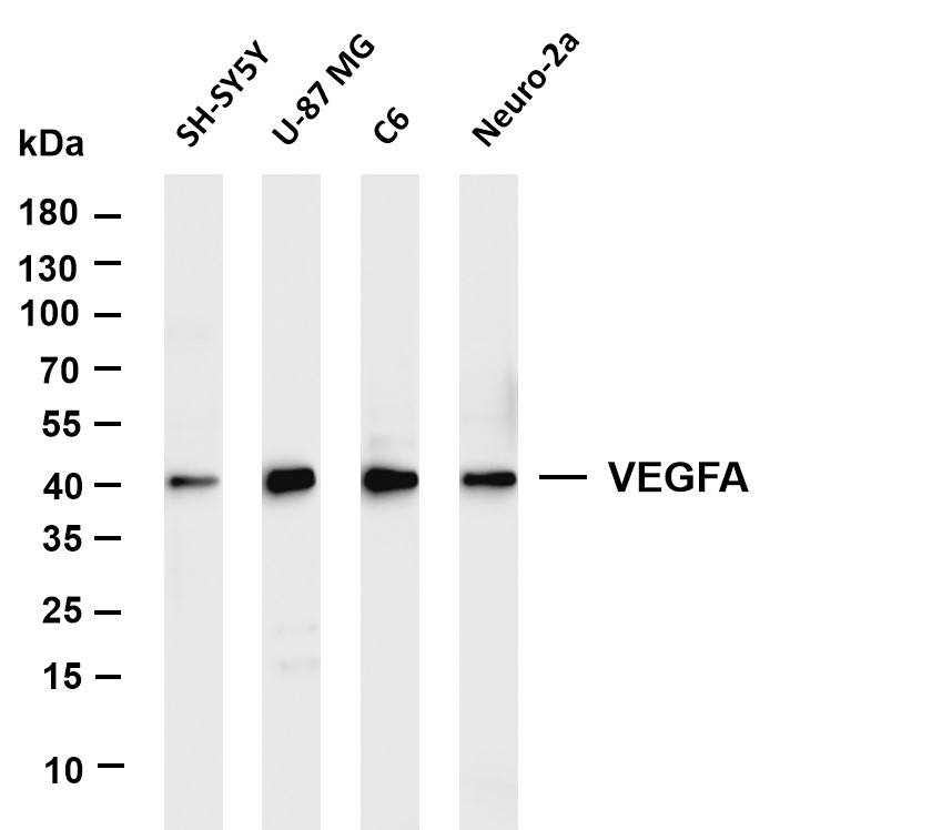
Various whole cell lysates were separated by 4-20% SDS-PAGE, and the membrane was blotted with anti-VEGFA antibody. The HRP-conjugated Goat anti-Rabbit IgG(H + L) antibody was used to detect the antibody. Lane 1: SH-SY5Y, Lane 2: U-87 MG, Lane 3: C6, Lane 4: Neuro-2a.IHC
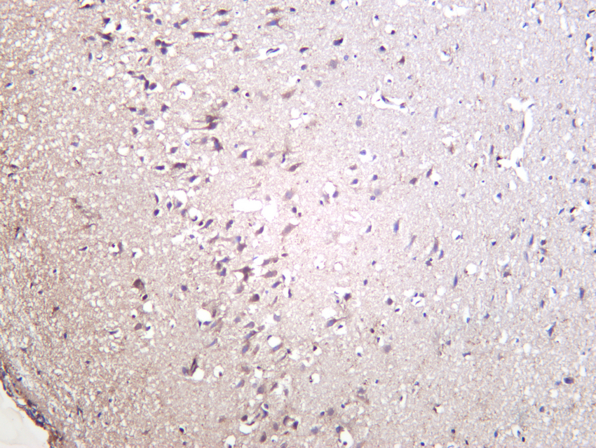
Rat brain was stained with anti-VEGFA rabbit antibody.IHC
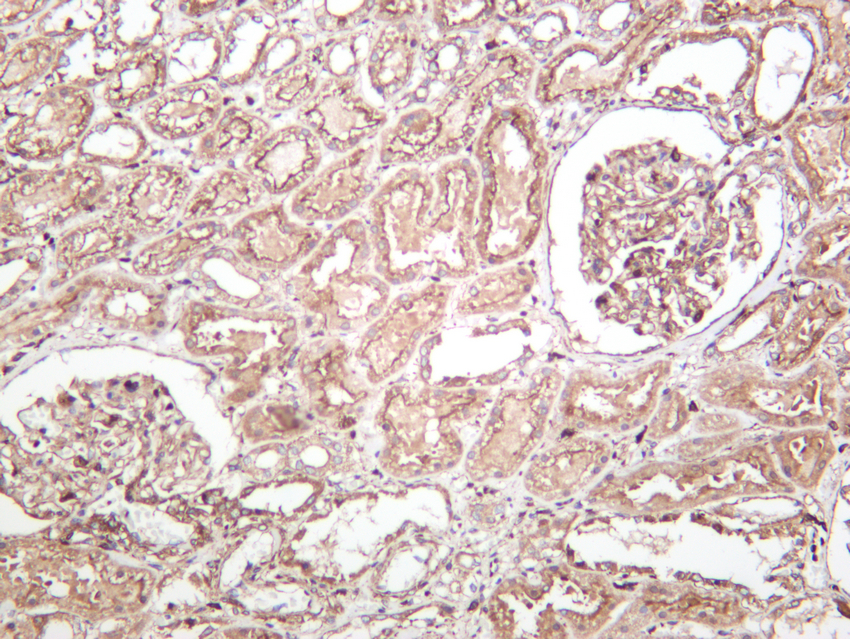
Human kidney was stained with anti-VEGFA rabbit antibody.IHC
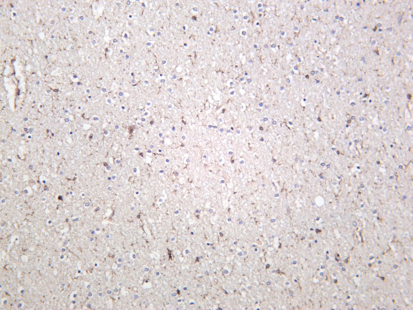
Mouse brain was stained with anti-VEGFA rabbit antibody.ICC/IF
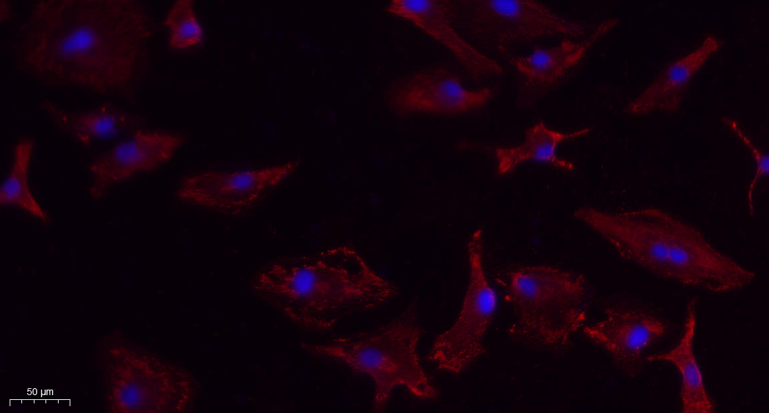
Immunofluorescence analysis of A549. 1,primary Antibody(red) was diluted at 1:200(4°C overnight). 2, Goat Anti Rabbit IgG (H&L) - Alexa Fluor 594 Secondary antibody was diluted at 1:1000(room temperature, 50min).3, DAPI(blue) 10min.| Product Name | VEGFA Rabbit mAb |
|---|---|
| Antibody Type | Primary Antibodies |
| Clonality | monoclonal |
|---|---|
| Isotype | IgG |
| Host Species | Rabbit |
| Tested Applications | ICC/IFIHCWB |
| WB:1:2000-1:10000 IHC:1:1000-1:5000 ICC/IF:1:200-1:1000 |
|
| Species Reactivity | HumanMouseRat |
| Concentration | 1mg/ml |
| Purification | Protein A |
| Gene Symbol | VEGFA |
|---|---|
| Gene Synonyms | VPF VEGF MVCD1 L-VEGF |
| Gene Full Name | vascular endothelial growth factor A |
| Gene Summary | This gene is a member of the PDGF/VEGF growth factor family. It encodes a heparin-binding protein, which exists as a disulfide-linked homodimer. This growth factor induces proliferation and migration of vascular endothelial cells, and is essential for both physiological and pathological angiogenesis. Disruption of this gene in mice resulted in abnormal embryonic blood vessel formation. This gene is upregulated in many known tumors and its expression is correlated with tumor stage and progression. Elevated levels of this protein are found in patients with POEMS syndrome, also known as Crow-Fukase syndrome. Allelic variants of this gene have been associated with microvascular complications of diabetes 1 (MVCD1) and atherosclerosis. Alternatively spliced transcript variants encoding different isoforms have been described. There is also evidence for alternative translation initiation from upstream non-AUG (CUG) codons resulting in additional isoforms. A recent study showed that a C-terminally extended isoform is produced by use of an alternative in-frame translation termination codon via a stop codon readthrough mechanism, and that this isoform is antiangiogenic. Expression of some isoforms derived from the AUG start codon is regulated by a small upstream open reading frame, which is located within an internal ribosome entry site. The levels of VEGF are increased during infection with severe acute respiratory syndrome coronavirus 2 (SARS-CoV-2), thus promoting inflammation by facilitating recruitment of inflammatory cells, and by increasing the level of angiopoietin II (Ang II), one of two products of the SARS-CoV-2 binding target, angiotensin-converting enzyme 2 (ACE2). In turn, Ang II facilitates the elevation of VEGF, thus forming a vicious cycle in the release of inflammatory cytokines. [provided by RefSeq, Jun 2020] |
| Alternative Names | VEGFA VEGF Vascular endothelial growth factor A VEGF-A Vascular permeability factor VPF |
| Molecular Weight(MW) | 27kDa(Observed band size: 40kDa) |
| Cellular Localization | Secreted. |
WB
Various whole cell lysates were separated by 4-20% SDS-PAGE, and the membrane was blotted with anti-VEGFA antibody. The HRP-conjugated Goat anti-Rabbit IgG(H + L) antibody was used to detect the antibody. Lane 1: SH-SY5Y, Lane 2: U-87 MG, Lane 3: C6, Lane 4: Neuro-2a.
IHC
Rat brain was stained with anti-VEGFA rabbit antibody.
IHC
Human kidney was stained with anti-VEGFA rabbit antibody.
IHC
Mouse brain was stained with anti-VEGFA rabbit antibody.
ICC/IF
Immunofluorescence analysis of A549. 1,primary Antibody(red) was diluted at 1:200(4°C overnight). 2, Goat Anti Rabbit IgG (H&L) - Alexa Fluor 594 Secondary antibody was diluted at 1:1000(room temperature, 50min).3, DAPI(blue) 10min.| Application Notes | WB:1:2000-1:10000 IHC:1:1000-1:5000 ICC/IF:1:200-1:1000 |
|---|
| Form | Liquid |
|---|---|
| Storage Instructions | -15°C to -25°C/1 year(Do not lower than -25°C) |
| Storage Buffer | PBS, 50% glycerol, 0.05% Proclin 300, 0.05%BSA. |


