WB
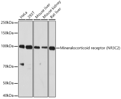
Western blot analysis of various lysates using Mineralocorticoid receptor (NR3C2) Rabbit pAb at 1:1000 dilution. Secondary antibody: HRP-conjugated Goat anti-Rabbit IgG (H+L) at 1:10000 dilution. Lysates/proteins: 25μg per lane. Blocking buffer: 3% nonfat dry milk in TBST. Detection: ECL Enhanced Kit. Exposure time: 90s.IHC
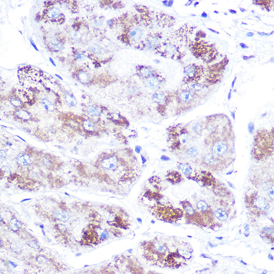
Immunohistochemistry analysis of paraffin embedded Human liver cancer using Mineralocorticoid receptor (NR3C2) Rabbit pAb at dilution of 1:100 (40x lens). High pressure antigen retrieval performed with 0.01M Citrate Bufferr (pH 6.0) prior to IHC staining.ICC/IF
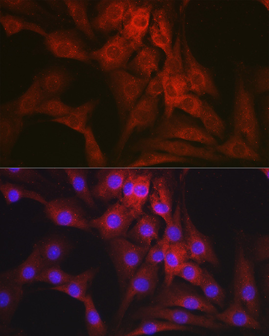
Immunofluorescence analysis of NIH-3T3 cells using Mineralocorticoid receptor (NR3C2) Rabbit pAb at dilution of 1:100 (40x lens). Secondary antibody: Cy3 conjugated Goat anti-Rabbit IgG (H+L) at 1:500 dilution. Blue: DAPI for nuclear staining.WB
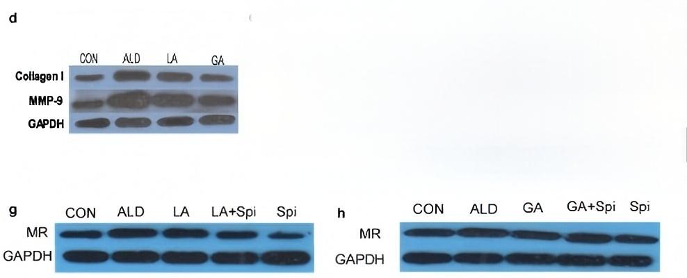
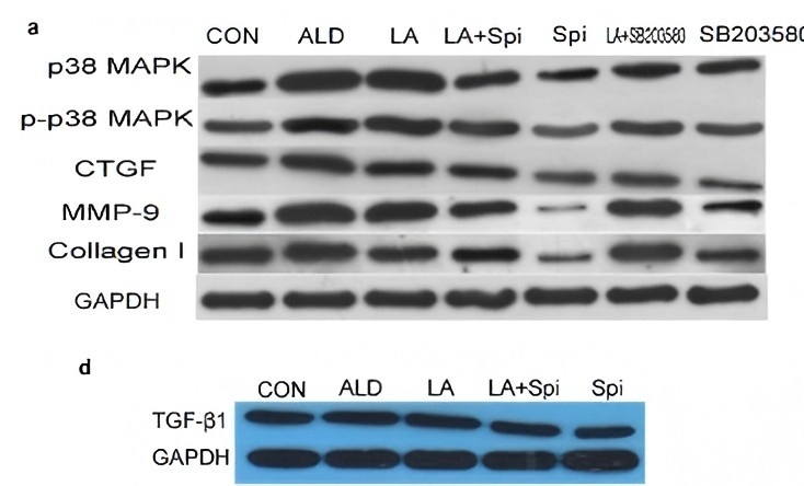
The protein lysates of H9c2 cells after treatment by ALD, LA, GA and Spi, respectively, were subjected to western blotting (d, g, h).WB
The protein lysates of H9c2 cells after treatment by LA, Spi and SB203580 administered alone or in combination were subjected to western blotting (a, d).| Product Name | Mineralocorticoid receptor (NR3C2) Rabbit pAb |
|---|---|
| Antibody Type | Primary Antibodies |
| Immunogen | Recombinant fusion protein containing a sequence corresponding to amino acids 450-600 of human Mineralocorticoid receptor (NR3C2) (NP_000892.2). |
| Clonality | Polyclonal |
|---|---|
| Isotype | IgG |
| Host Species | Rabbit |
| Tested Applications | ICC/IFIHCWB |
| WB:1:500-1:1000 IHC:1:50-1:200 ICC/IF:1:50-1:200 |
|
| Species Reactivity | HumanMouseRat |
| Concentration | 1mg/ml |
| Purification | Affinity purified |
| Gene Symbol | NR3C2 |
|---|---|
| Gene Synonyms | MR MCR MLR NR3C2VIT |
| Gene Full Name | nuclear receptor subfamily 3 group C member 2 |
| Gene Summary | This gene encodes the mineralocorticoid receptor, which mediates aldosterone actions on salt and water balance within restricted target cells. The protein functions as a ligand-dependent transcription factor that binds to mineralocorticoid response elements in order to transactivate target genes. Mutations in this gene cause autosomal dominant pseudohypoaldosteronism type I, a disorder characterized by urinary salt wasting. Defects in this gene are also associated with early onset hypertension with severe exacerbation in pregnancy. Alternative splicing results in multiple transcript variants. [provided by RefSeq, Oct 2009] |
| Alternative Names | MR MCR MLR NR3C2VIT Mineralocorticoid receptor (NR3C2) |
| Molecular Weight(MW) | 107kDa(Observed MW 100kDa) |
| Cellular Localization | Cytoplasm, Endoplasmic reticulum membrane, Nucleus, Peripheral membrane protein. |
WB
Western blot analysis of various lysates using Mineralocorticoid receptor (NR3C2) Rabbit pAb at 1:1000 dilution. Secondary antibody: HRP-conjugated Goat anti-Rabbit IgG (H+L) at 1:10000 dilution. Lysates/proteins: 25μg per lane. Blocking buffer: 3% nonfat dry milk in TBST. Detection: ECL Enhanced Kit. Exposure time: 90s.
IHC
Immunohistochemistry analysis of paraffin embedded Human liver cancer using Mineralocorticoid receptor (NR3C2) Rabbit pAb at dilution of 1:100 (40x lens). High pressure antigen retrieval performed with 0.01M Citrate Bufferr (pH 6.0) prior to IHC staining.
ICC/IF
Immunofluorescence analysis of NIH-3T3 cells using Mineralocorticoid receptor (NR3C2) Rabbit pAb at dilution of 1:100 (40x lens). Secondary antibody: Cy3 conjugated Goat anti-Rabbit IgG (H+L) at 1:500 dilution. Blue: DAPI for nuclear staining.
WB
The protein lysates of H9c2 cells after treatment by ALD, LA, GA and Spi, respectively, were subjected to western blotting (d, g, h).
WB
The protein lysates of H9c2 cells after treatment by LA, Spi and SB203580 administered alone or in combination were subjected to western blotting (a, d).| Application Notes | WB:1:500-1:1000 IHC:1:50-1:200 ICC/IF:1:50-1:200 |
|---|
| Form | Liquid |
|---|---|
| Storage Instructions | Store at -20℃. Avoid freeze / thaw cycles. |
| Storage Buffer | PBS with 0.01% thimerosal, 50% glycerol, pH7.3. |


