WB
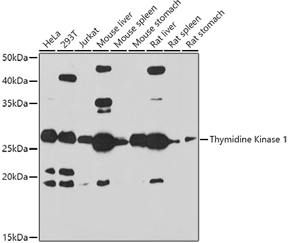
Western blot analysis of various lysates using Thymidine Kinase 1 Rabbit pAb at 1:1000 dilution. Secondary antibody: HRP-conjugated Goat anti-Rabbit IgG (H+L) at 1:10000 dilution. Lysates/proteins: 25μg per lane. Blocking buffer: 3% nonfat dry milk in TBST. Detection: ECL Basic Kit.Exposure time:180s.IHC
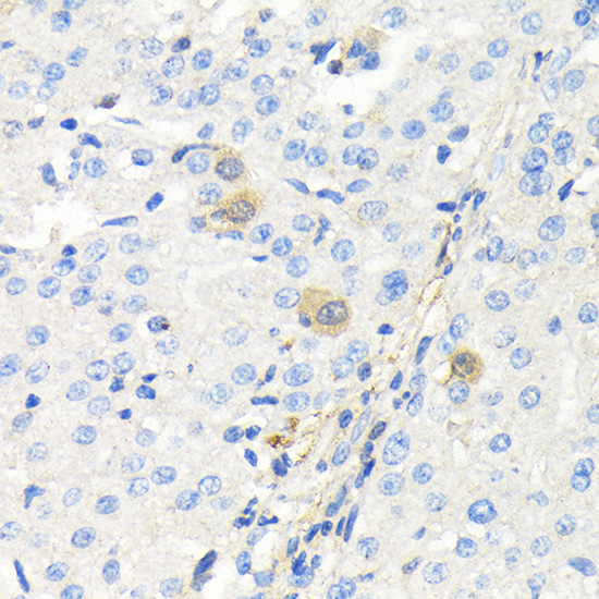
Immunohistochemistry analysis of paraffin embedded Human liver cancer using Thymidine Kinase 1 Rabbit pAb at dilution of 1:100 (40x lens). Microwave antigen retrieval performed with 0.01M Tris/EDTA Buffer (pH 9.0) prior to IHC staining.ICC/IF
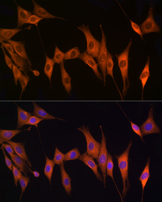
Immunofluorescence analysis of NIH/3T3 cells using Thymidine Kinase 1 Rabbit pAb at dilution of 1:100 (40x lens). Secondary antibody: Cy3-conjugated Goat anti-Rabbit IgG (H+L) at 1:500 dilution. Blue: DAPI for nuclear staining.IF
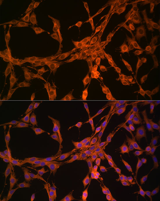
Immunofluorescence analysis of PC-12 cells using Thymidine Kinase 1 Rabbit pAb at dilution of 1:100 (40x lens). Secondary antibody: Cy3-conjugated Goat anti-Rabbit IgG (H+L) at 1:500 dilution. Blue: DAPI for nuclear staining.| Product Name | Thymidine Kinase 1 Rabbit pAb |
|---|---|
| Antibody Type | Primary Antibodies |
| Immunogen | Recombinant fusion protein containing a sequence corresponding to amino acids 15-194 of human Thymidine Kinase 1 (NP_003249.3). |
| Clonality | Polyclonal |
|---|---|
| Isotype | IgG |
| Host Species | Rabbit |
| Tested Applications | ICC/IFIHCWB |
| WB:1:500-1:1000 IHC:1:50-1:200 ICC/IF:1:50-1:200 |
|
| Species Reactivity | HumanMouseRat |
| Concentration | 1mg/ml |
| Purification | Affinity purified |
| Gene Symbol | TK1 |
|---|---|
| Gene Synonyms | TK2 |
| Gene Full Name | thymidine kinase 1 |
| Gene Summary | The protein encoded by this gene is a cytosolic enzyme that catalyzes the addition of a gamma-phosphate group to thymidine. This creates dTMP and is the first step in the biosynthesis of dTTP, which is one component required for DNA replication. The encoded protein, whose levels fluctuate depending on the cell cycle stage, can act as a low activity dimer or a high activity tetramer. High levels of this protein have been used as a biomarker for diagnosing and categorizing many types of cancers. [provided by RefSeq, Oct 2016] |
| Alternative Names | TK2 Thymidine Kinase 1 |
| Molecular Weight(MW) | 26kDa |
| Cellular Localization | Cytoplasm. |
WB
Western blot analysis of various lysates using Thymidine Kinase 1 Rabbit pAb at 1:1000 dilution. Secondary antibody: HRP-conjugated Goat anti-Rabbit IgG (H+L) at 1:10000 dilution. Lysates/proteins: 25μg per lane. Blocking buffer: 3% nonfat dry milk in TBST. Detection: ECL Basic Kit.Exposure time:180s.
IHC
Immunohistochemistry analysis of paraffin embedded Human liver cancer using Thymidine Kinase 1 Rabbit pAb at dilution of 1:100 (40x lens). Microwave antigen retrieval performed with 0.01M Tris/EDTA Buffer (pH 9.0) prior to IHC staining.
ICC/IF
Immunofluorescence analysis of NIH/3T3 cells using Thymidine Kinase 1 Rabbit pAb at dilution of 1:100 (40x lens). Secondary antibody: Cy3-conjugated Goat anti-Rabbit IgG (H+L) at 1:500 dilution. Blue: DAPI for nuclear staining.
IF
Immunofluorescence analysis of PC-12 cells using Thymidine Kinase 1 Rabbit pAb at dilution of 1:100 (40x lens). Secondary antibody: Cy3-conjugated Goat anti-Rabbit IgG (H+L) at 1:500 dilution. Blue: DAPI for nuclear staining.| Application Notes | WB:1:500-1:1000 IHC:1:50-1:200 ICC/IF:1:50-1:200 |
|---|
| Form | Liquid |
|---|---|
| Storage Instructions | Store at -20℃. Avoid freeze / thaw cycles. |
| Storage Buffer | PBS with 0.09% Sodium azide, 50% glycerol, pH7.3. |


