WB
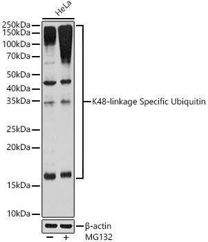
Western blot analysis of lysates from HeLa cells using K48-linkage Specific Ubiquitin Rabbit mAb at 1:1000 dilution incubated overnight at 4℃. HeLa cells were treated by MG132 (50 μM) at 37℃ for 90 minutes. Secondary antibody: HRP-conjugated Goat anti-Rabbit IgG (H+L) at 1:10000 dilution. Lysates/proteins: 25μg per lane. Blocking buffer: 3% nonfat dry milk in TBST. Detection: ECL Basic Kit. Exposure time: 180s.ICC/IF
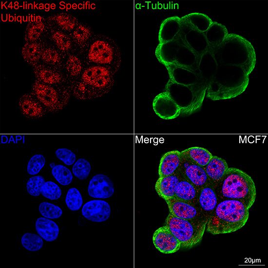
Confocal imaging of MCF7 cells using K48 linkage Specific Ubiquitin Rabbit mAb (dilution 1:100) followed by a further incubation with Cy3 Goat Anti-Rabbit IgG (H+L) (dilution 1:500)(Red).The cells were counterstained with α-Tubulin Mouse mAb (dilution 1:400) followed by incubation with A Omnimabs® 488 Goat Anti-Mouse IgG(H&L) Ab (dilution 1:500) (Green).DAPI was used for nuclear staining (Blue). Objective: 100x.ICC/IF
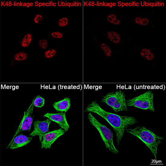
Confocal imaging of HeLa cells (treated with MG-132) and HeLa cells (untreated) cells using K48-linkage Specific Ubiquitin Rabbit mAb (dilution 1:200) followed by a further incubation with Cy3 Goat Anti-Rabbit IgG (H+L) (dilution 1:500) (Red). The cells were counterstained with α-Tubulin Mouse mAb (dilution 1:400) followed by incubation with Omnimabs® 488 Goat Anti-Mouse IgG(H&L) Ab (dilution 1:500) (Green). DAPI was used for nuclear staining (Blue). Objective: 100x.| Product Name | K48-linkage Specific Ubiquitin Rabbit mAb |
|---|---|
| Antibody Type | Primary Antibodies |
| Immunogen | A synthetic peptide corresponding to a sequence containing Ubiquitin protein (K48 linkage). |
| Clonality | Monoclonal |
|---|---|
| Isotype | IgG |
| Host Species | Rabbit |
| Tested Applications | ICC/IFWB |
| WB:1:500-1:2000 ICC/IF:1:50-1:200 |
|
| Species Reactivity | HumanMouse |
| Concentration | 1mg/ml |
| Purification | Affinity purified |
| Gene Symbol | UBB |
|---|---|
| Gene Synonyms | HEL-S-50 |
| Gene Full Name | ubiquitin B |
| Gene Summary | This gene encodes ubiquitin, one of the most conserved proteins known. Ubiquitin has a major role in targeting cellular proteins for degradation by the 26S proteosome. It is also involved in the maintenance of chromatin structure, the regulation of gene expression, and the stress response. Ubiquitin is synthesized as a precursor protein consisting of either polyubiquitin chains or a single ubiquitin moiety fused to an unrelated protein. This gene consists of three direct repeats of the ubiquitin coding sequence with no spacer sequence. Consequently, the protein is expressed as a polyubiquitin precursor with a final amino acid after the last repeat. An aberrant form of this protein has been detected in patients with Alzheimer's disease and Down syndrome. Pseudogenes of this gene are located on chromosomes 1, 2, 13, and 17. Alternative splicing results in multiple transcript variants. [provided by RefSeq, Aug 2013] |
| Alternative Names | HEL-S-50 K48-linkage Specific Ubiquitin |
| Molecular Weight(MW) | 26kDa(Observed MW:17-250kDa) |
| Cellular Localization | Cytoplasm, Nucleus. |
WB
Western blot analysis of lysates from HeLa cells using K48-linkage Specific Ubiquitin Rabbit mAb at 1:1000 dilution incubated overnight at 4℃. HeLa cells were treated by MG132 (50 μM) at 37℃ for 90 minutes. Secondary antibody: HRP-conjugated Goat anti-Rabbit IgG (H+L) at 1:10000 dilution. Lysates/proteins: 25μg per lane. Blocking buffer: 3% nonfat dry milk in TBST. Detection: ECL Basic Kit. Exposure time: 180s.
ICC/IF
Confocal imaging of MCF7 cells using K48 linkage Specific Ubiquitin Rabbit mAb (dilution 1:100) followed by a further incubation with Cy3 Goat Anti-Rabbit IgG (H+L) (dilution 1:500)(Red).The cells were counterstained with α-Tubulin Mouse mAb (dilution 1:400) followed by incubation with A Omnimabs® 488 Goat Anti-Mouse IgG(H&L) Ab (dilution 1:500) (Green).DAPI was used for nuclear staining (Blue). Objective: 100x.
ICC/IF
Confocal imaging of HeLa cells (treated with MG-132) and HeLa cells (untreated) cells using K48-linkage Specific Ubiquitin Rabbit mAb (dilution 1:200) followed by a further incubation with Cy3 Goat Anti-Rabbit IgG (H+L) (dilution 1:500) (Red). The cells were counterstained with α-Tubulin Mouse mAb (dilution 1:400) followed by incubation with Omnimabs® 488 Goat Anti-Mouse IgG(H&L) Ab (dilution 1:500) (Green). DAPI was used for nuclear staining (Blue). Objective: 100x.| Application Notes | WB:1:500-1:2000 ICC/IF:1:50-1:200 |
|---|
| Form | Liquid |
|---|---|
| Storage Instructions | Store at -20℃. Avoid freeze / thaw cycles. |
| Storage Buffer | PBS with 0.02% sodium azide, 0.05% BSA, 50% glycerol, pH7.3. |


