WB
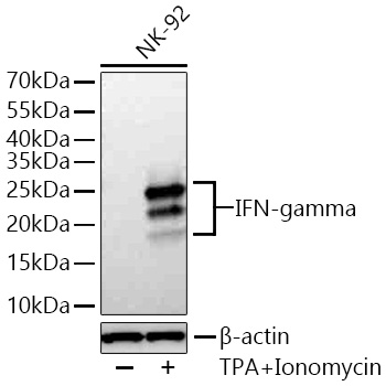
Western blot analysis of lysates from NK-92 cells using IFN-gamma Rabbit mAb at 1:5000 dilution incubated overnight at 4℃. NK-92 cells were treated by TPA (80 nM) and Ionomycin (3 μM) at 37℃ for 4 hours after serum-starvation overnight. Secondary antibody: HRP-conjugated Goat anti-Rabbit IgG (H+L) at 1:10000 dilution. Lysates/proteins: 30 μg per lane. Blocking buffer: 3% nonfat dry milk in TBST. Detection: ECL Basic Kit. Exposure time: 45s.ICC/IF
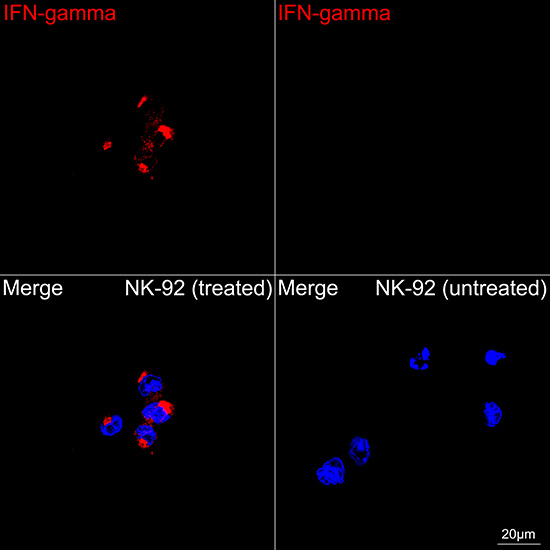
Confocal imaging of NK-92 cells (treated with TPA and Ionomycin) and NK-92 cells (untreated) using IFN-gamma Rabbit mAb (dilution 1:300) followed by a further incubation with Cy3 Goat Anti-Rabbit IgG (H+L) (dilution 1:500) (Red). DAPI was used for nuclear staining (Blue). Objective: 100x.IP
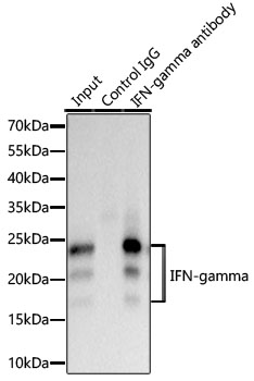
Immunoprecipitation of IFN-gamma from 600 µg extracts of NK-92 cells treated by TPA 80nM and Ionomycin 3μM for 4 hours was performed using 0.5 µg of IFN-gamma Rabbit mAb. Rabbit IgG isotype control was used to precipitate the Control IgG sample. IP samples were eluted with 1X reducing Laemmli Buffer. The Input lane represents 10% of the total input. Western blot analysis of immunoprecipitates was conducted using IFN-gamma Rabbit mAb at a dilution of 1:2000.| Product Name | IFN-gamma Rabbit mAb |
|---|---|
| Antibody Type | Primary Antibodies |
| Immunogen | Recombinant fusion protein containing a sequence corresponding to amino acids 24-166 of human IFN-gamma (NP_000610.2). |
| Clonality | Monoclonal |
|---|---|
| Isotype | IgG |
| Host Species | Rabbit |
| Tested Applications | ICC/IFIPWB |
| WB:1:5000-1:30000 ICC/IF:1:200-1:600 IP:0.5μg-4μg antibody for 500μg-700μg extracts of whole cells |
|
| Species Reactivity | Human |
| Concentration | 1mg/ml |
| Purification | Affinity purified |
| Gene Symbol | IFNG |
|---|---|
| Gene Synonyms | IFG IFI IMD69 |
| Gene Full Name | interferon gamma |
| Gene Summary | This gene encodes a soluble cytokine that is a member of the type II interferon class. The encoded protein is secreted by cells of both the innate and adaptive immune systems. The active protein is a homodimer that binds to the interferon gamma receptor which triggers a cellular response to viral and microbial infections. Mutations in this gene are associated with an increased susceptibility to viral, bacterial and parasitic infections and to several autoimmune diseases. [provided by RefSeq, Dec 2015] |
| Alternative Names | IFG IFI IMD69 |
| Molecular Weight(MW) | 19kDa(Observed MW:17kDa/19kDa/23kDa) |
| Cellular Localization | Secreted. |
WB
Western blot analysis of lysates from NK-92 cells using IFN-gamma Rabbit mAb at 1:5000 dilution incubated overnight at 4℃. NK-92 cells were treated by TPA (80 nM) and Ionomycin (3 μM) at 37℃ for 4 hours after serum-starvation overnight. Secondary antibody: HRP-conjugated Goat anti-Rabbit IgG (H+L) at 1:10000 dilution. Lysates/proteins: 30 μg per lane. Blocking buffer: 3% nonfat dry milk in TBST. Detection: ECL Basic Kit. Exposure time: 45s.
ICC/IF
Confocal imaging of NK-92 cells (treated with TPA and Ionomycin) and NK-92 cells (untreated) using IFN-gamma Rabbit mAb (dilution 1:300) followed by a further incubation with Cy3 Goat Anti-Rabbit IgG (H+L) (dilution 1:500) (Red). DAPI was used for nuclear staining (Blue). Objective: 100x.
IP
Immunoprecipitation of IFN-gamma from 600 µg extracts of NK-92 cells treated by TPA 80nM and Ionomycin 3μM for 4 hours was performed using 0.5 µg of IFN-gamma Rabbit mAb. Rabbit IgG isotype control was used to precipitate the Control IgG sample. IP samples were eluted with 1X reducing Laemmli Buffer. The Input lane represents 10% of the total input. Western blot analysis of immunoprecipitates was conducted using IFN-gamma Rabbit mAb at a dilution of 1:2000.| Application Notes | WB:1:5000-1:30000 ICC/IF:1:200-1:600 IP:0.5μg-4μg antibody for 500μg-700μg extracts of whole cells |
|---|
| Form | Liquid |
|---|---|
| Storage Instructions | Store at -20℃. Avoid freeze / thaw cycles. |
| Storage Buffer | PBS with 0.09% Sodium azide, 0.05% BSA, 50% glycerol, pH7.3. |


