WB
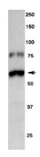
Western blot showing specific immunolabeling of tyrosine hydroxylase.IHC
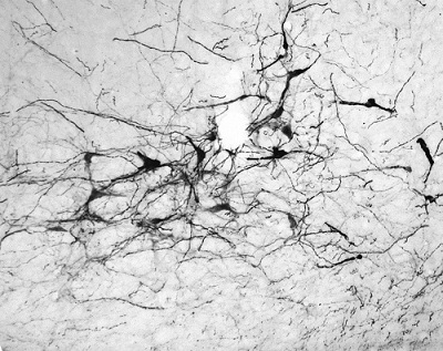
Immunohistochemistry (Free Floating) analysis of the rostral ventral lateral medulla in adult mouse brain labelling tyrosine hydroxylase with Tyrosine Hydroxylase antibody at a dilution of 1/1000. Tissue was fixed with 10% formalin.IF-P
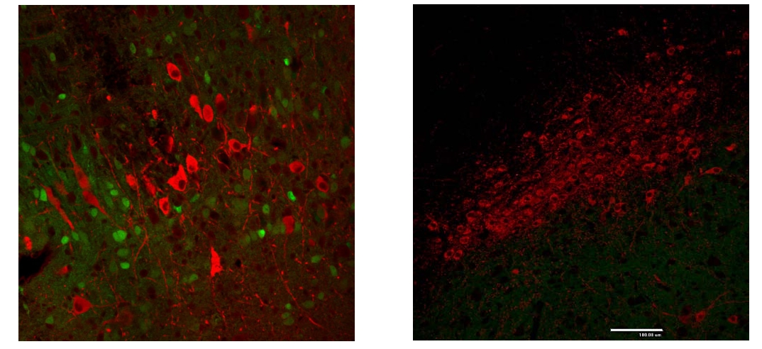
Immunostaining of paraformaldehyde-fixed (4%) paraffin-embedded sections of the substantia nigra pars compacta of an adult mouse brain. This mouse was engineered to have GFP expressed under control of the actin promoter, which explains the low-level green autofluorescence. Note the high number of dopaminergic neuron cell bodies in this brain region. Detection of the chicken antibody came from a Texas Red-labeled goat anti-chicken IgY, 1:500.| Product Name | Anti-Tyrosine Hydroxylase (TYH) Antibody |
|---|---|
| Antibody Type | Primary Antibodies |
| Immunogen | Two synthetic peptide/ keyhole limpet hemocyanin (KLH) conjugates. These synthetic peptides corresponded to different regions of the Tyrosine Hydroxylase gene product, but were shared between the human (P07101) and mouse (P24529) sequences. |
| Clonality | Polyclonal |
|---|---|
| Isotype | IgY |
| Host Species | Chicken |
| Tested Applications | IF-PIHCWB |
| WB:1:1000-1:2000 IHC:1:1000-1:2000 IF-P:1:1000-1:2000 |
|
| Species Reactivity | HumanMouseRat |
| Concentration | 0.2mg/ml |
| Purification | Affinity purified |
| Target Description | Human TYH (EC 1.14.16.2) is a 58,523 dalton protein (528 amino acids) responsible for the enzymatic conversion of L-tyrosine to L-DOPA (dihydroxyphenylalanine). This enzyme is expressed in all catecholaminergic neurons of the CNS and PNS. In the CNS, TYH-positive neurons can be found within the substantia nigra, ventral tegmental area, locus ceruleus, and hypothalamus. In the PNS, TYH-positive neurons can be found within the sympathetic chain, pre-vertebral ganglia and the adrenal medulla. |
|---|---|
| Alternative Names | Tyrosine 3-monooxygenase (EC 1.14.16.2) (Tyrosine 3-hydroxylase) (TH) |
| Molecular Weight(MW) | 60 kDa |
WB
Western blot showing specific immunolabeling of tyrosine hydroxylase.
IHC
Immunohistochemistry (Free Floating) analysis of the rostral ventral lateral medulla in adult mouse brain labelling tyrosine hydroxylase with Tyrosine Hydroxylase antibody at a dilution of 1/1000. Tissue was fixed with 10% formalin.
IF-P
Immunostaining of paraformaldehyde-fixed (4%) paraffin-embedded sections of the substantia nigra pars compacta of an adult mouse brain. This mouse was engineered to have GFP expressed under control of the actin promoter, which explains the low-level green autofluorescence. Note the high number of dopaminergic neuron cell bodies in this brain region. Detection of the chicken antibody came from a Texas Red-labeled goat anti-chicken IgY, 1:500.| Application Notes | WB:1:1000-1:2000 IHC:1:1000-1:2000 IF-P:1:1000-1:2000 |
|---|
| Form | Liquid |
|---|---|
| Storage Instructions | Store at 4°C in the dark. Under these conditions, the antibodies should have a shelf life of at least twelve months, provided they remain sterile. For longer term storage, aliquot and freeze to avoid freeze-thaw of the antibody. |
| Storage Buffer | Phosphate-buffered (10 mM) isotonic (0.9%, w/v) saline (“PBS," pH 7.2) with sodium azide (0.02%, w/v) added as a preservative. |


