WB
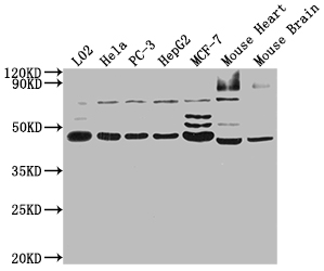
Positive WB detected in: L02 whole cell lysate, Hela whole cell lysate, PC-3 whole cell lysate, HepG2 whole cell lysate, MCF-7 whole cell lysate, Mouse Heart tissue, Mouse Brain tissue. All lanes: NOB1 antibody at 1:2000, Secondary Goat polyclonal to rabbit IgG at 1/50000 dilution.IHC
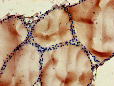
Immunohistochemistry of paraffin-embedded human thyroid tissue using NOB1 Antibody at dilution of 1:100.IHC
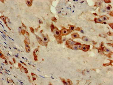
Immunohistochemistry of paraffin-embedded human placenta tissue using NOB1 Antibody at dilution of 1:100.ICC/IF
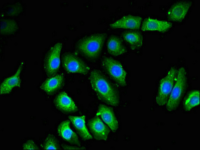
Immunofluorescent analysis of A549 cells using NOB1 Antibody at dilution of 1:100 and Alexa Fluor 488-congugated AffiniPure Goat Anti-Rabbit IgG(H+L).| Product Name | Rabbit anti-NOB1 Polyclonal antibody |
|---|---|
| Antibody Type | Primary Antibodies |
| Immunogen | Recombinant Human RNA-binding protein NOB1 protein (103-229AA). |
| Clonality | Polyclonal |
|---|---|
| Isotype | IgG |
| Host Species | Rabbit |
| Tested Applications | ICC/IFIHCWB |
| WB:1:500-1:5000 IHC:1:20-1:200 ICC/IF:1:50-1:200 |
|
| Species Reactivity | HumanMouse |
| Concentration | 1mg/ml |
| Purification | Protein G |
| Gene Symbol | NOB1 |
|---|---|
| Gene Synonyms | ART-4 NOB1P MST158 MSTP158 PSMD8BP1 |
| Gene Full Name | NIN1 (RPN12) binding protein 1 homolog |
| Gene Summary | In yeast, over 200 protein and RNA cofactors are required for ribosome assembly, and these are generally conserved in eukaryotes. These factors orchestrate modification and cleavage of the initial 35S precursor rRNA transcript into the mature 18S, 5.8S, and 25S rRNAs, folding of the rRNA, and binding of ribosomal proteins and 5S RNA. Nob1 is involved in pre-rRNA processing. In a late cytoplasmic processing step, Nob1 cleaves a 20S rRNA intermediate at cleavage site D to produce the mature 18S rRNA (Lamanna and Karbstein, 2009 [PubMed 19706509]).[supplied by OMIM, Nov 2010] |
| Alternative Names | Adenocarcinoma antigen recognized by T lymphocytes 4 antibody ART 4 antibody ART4 antibody MST158 antibody MSTP158 antibody Nin one binding protein antibody NIN1/PSMD8 binding protein 1 homolog antibody NIN1/RPN12 binding protein 1 homolog (S. cerevisiae) antibody nob1 antibody NOB1_HUMAN antibody NOB1P antibody Phosphorylation regulatory protein HP-10 antibody Protein ART-4 antibody Protein ART4 antibody PSMD8 binding protein 1 antibody PSMD8BP1 antibody RNA binding protein NOB1 antibody RNA-binding protein NOB1 antibody |
| Molecular Weight(MW) | 47kDa |
| Cellular Localization | Nucleus. |
WB
Positive WB detected in: L02 whole cell lysate, Hela whole cell lysate, PC-3 whole cell lysate, HepG2 whole cell lysate, MCF-7 whole cell lysate, Mouse Heart tissue, Mouse Brain tissue. All lanes: NOB1 antibody at 1:2000, Secondary Goat polyclonal to rabbit IgG at 1/50000 dilution.
IHC
Immunohistochemistry of paraffin-embedded human thyroid tissue using NOB1 Antibody at dilution of 1:100.
IHC
Immunohistochemistry of paraffin-embedded human placenta tissue using NOB1 Antibody at dilution of 1:100.
ICC/IF
Immunofluorescent analysis of A549 cells using NOB1 Antibody at dilution of 1:100 and Alexa Fluor 488-congugated AffiniPure Goat Anti-Rabbit IgG(H+L).| Application Notes | WB:1:500-1:5000 IHC:1:20-1:200 ICC/IF:1:50-1:200 |
|---|
| Form | Liquid |
|---|---|
| Storage Instructions | Upon receipt, store at -20°C or -80°C. Avoid repeated freeze. |
| Storage Buffer | Preservative: 0.03% Proclin 300,Constituents: 50% Glycerol, 0.01M PBS, pH 7.4 |


